. 2008 Feb 12;105(6):1861-6.
doi: 10.1073/pnas.0705753105. Epub 2008 Feb 5.

Structure and mechanism of IFN-gamma antagonism by an orthopoxvirus IFN-gamma-binding protein

Affiliations

Structure and mechanism of IFN-gamma antagonism by an orthopoxvirus IFN-gamma-binding protein

Anthony A Nuara et al. Proc Natl Acad Sci U S A. .

Abstract

Ectromelia virus (ECTV) encodes an IFN-gamma-binding protein (IFN-gammaBP(ECTV)) that disrupts IFN-gamma signaling and its ability to induce an antiviral state within cells. IFN-gammaBP(ECTV) is an important virulence factor that is highly conserved (>90%) in all orthopoxviruses, including variola virus, the causative agent of smallpox. The 2.2-A crystal structure of the IFN-gammaBP(ECTV)/IFN-gamma complex reveals IFN-gammaBP(ECTV) consists of an IFN-gammaR1 ligand-binding domain and a 57-aa helix-turn-helix (HTH) motif that is structurally related to the transcription factor TFIIA. The HTH motif forms a tetramerization domain that results in an IFN-gammaBP(ECTV)/IFN-gamma complex containing four IFN-gammaBP(ECTV) chains and two IFN-gamma dimers. The structure, combined with biochemical and cell-based assays, demonstrates that IFN-gammaBP(ECTV) tetramers are required for efficient IFN-gamma antagonism.


Conflict of interest statement

The authors declare no conflict of interest.

Figures

Fig. 1.
Fig. 1.
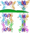
Structure of IFN-γBPECTV. (A) Ribbon diagram of the IFN-γBPECTV monomer. (B) Ribbon diagram of the IFN-γBPECTV dimer. Chains R and T are colored cyan and magenta, respectively. (C) Close-up of the T/R dimer interface (boxed region in B). The molecular surface colored by residue type (D and E, red; K and R, blue; G, magenta; C, yellow; S, T, H, N, and Q, orange; A, P, V, M, L, I, F, and W, green) is shown for chain R. Protruding residues in the EF and CC′ loops of the IFN-γR1 that prevent “IFN-γBPECTV-like” dimerization are shown in stick representation. (D) Ribbon diagram of the IFN-γBPECTV tetramer.
Fig. 2.
Fig. 2.
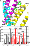
Structure and function of the HTH tetramer. (A) Close-up of R/T HTH dimer (chains R and T) forming one side of the tetramer interaction and HTH chain S colored as described in Fig. 1. (B) Surface area buried by each HTH residue in the IFN-γBPECTV dimer (black bars) and tetramer interfaces (red bars). The single alanine mutants tested in this study are circled. The labels for some of the alanine mutants analyzed in other work [residues 258–266 (9)] are boxed. Of the 14 mutants tested, only the Phe250Ala mutation (marked by an asterisk) significantly disrupts IFN-γ neutralization and IFN-γBPECTV tetramer formation (see SI Figs. 7 and 8).
Fig. 3.
Fig. 3.
Comparison of IFN-γ/IFN-γR1 and IFN-γ/ IFN-γBPECTV complexes. (A–D) Orthogonal views of the IFN-γ/IFN-γR1 (A and B) and IFN-γ/ IFN-γBPECTV (C and D) complexes.
Fig. 4.
Fig. 4.
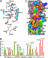
The IFN-γ/IFN-γBPECTV-binding interface. (A) Ribbon diagram of IFN-γ residues that contact IFN-γBPECTV. The residues that bury surface area into the interface are colored red. (B) Comparison of surface area buried by IFN-γ residues in the IFN-γ/IFN-γBPECTV (red) or IFN-γ/IFN-γR1 (green) complexes. (C) IFN-γBPECTV residues that bury surface area in the interface (≤3 Å2, gray; >3 to ≤10 Å2, blue; >10 to ≤20 Å2, cyan; >20 to ≤40 Å2, green; >40 to ≤60 Å2, yellow; >60 to ≤80 Å2, orange; >80 Å2, red). IFN-γ is shown as a magenta ribbon, with the side chains shown in A.
Fig. 5.
Fig. 5.
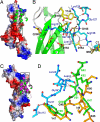
IFN-γ C terminus and AB loop interactions with IFN-γBPECTV and IFN-γR1. (A) Electrostatic surface potential of IFN-γBPECTV and the IFN-γ-binding epitope. (B) Detailed interactions between the C terminus of IFN-γ (cyan) and IFN-γBPECTV (yellow) corresponding to the box in A. (C) Electrostatic surface potential of IFN-γR1 in the same orientation as IFN-γBPECTV in A with IFN-γ from the IFN-γBPECTV (green) and IFN-γR1 (magenta) complexes. (D) Hydrogen bonding in the AB loop regions (boxed region in C) of the complexes. IFN-γ side chains are cyan, whereas L2 loop residues from IFN-γBPECTV and IFN-γR1 are colored gold and green, respectively. Gly-26* denotes the position of Gly-26IFN-γ, which is deleted in the AB loop of murine IFN-γ.

References

    1. Esteban DJ, Buller RM. Ectromelia virus: The causative agent of mousepox. J Gen Virol. 2005;86:2645–2659. - PubMed
    1. Seet BT, et al. Poxviruses and immune evasion. Annu Rev Immunol. 2003;21:377–423. - PubMed
    1. Alcami A. Viral mimicry of cytokines, chemokines and their receptors. Nat Rev Immunol. 2003;3:36–50. - PubMed
    1. Karupiah G, et al. Importance of interferons in recovery from mousepox. J Virol. 1993;67:4214–4226. - PMC - PubMed
    1. Chaudhri G, et al. Polarized type 1 cytokine response and cell-mediated immunity determine genetic resistance to mousepox. Proc Natl Acad Sci USA. 2004;101:9057–9062. - PMC - PubMed

Publication types

Associated data

LinkOut - more resources