. 2016 May 23;213(4):479-94.
doi: 10.1083/jcb.201510065.

Extracellular chloride signals collagen IV network assembly during basement membrane formation

Affiliations

Extracellular chloride signals collagen IV network assembly during basement membrane formation

Christopher F Cummings et al. J Cell Biol. .

Abstract

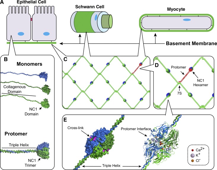
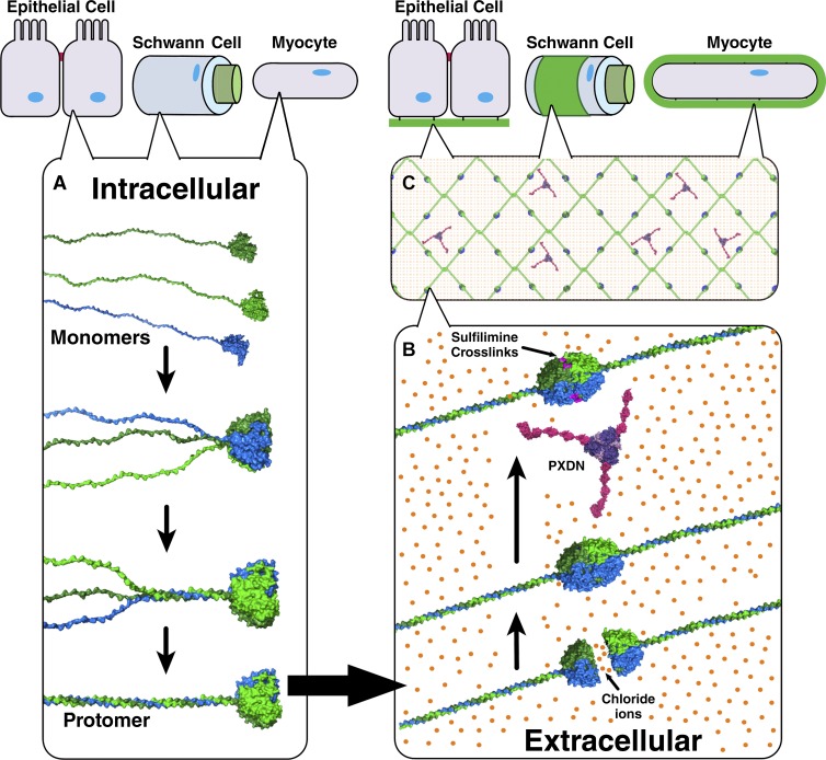
Basement membranes are defining features of the cellular microenvironment; however, little is known regarding their assembly outside cells. We report that extracellular Cl(-) ions signal the assembly of collagen IV networks outside cells by triggering a conformational switch within collagen IV noncollagenous 1 (NC1) domains. Depletion of Cl(-) in cell culture perturbed collagen IV networks, disrupted matrix architecture, and repositioned basement membrane proteins. Phylogenetic evidence indicates this conformational switch is a fundamental mechanism of collagen IV network assembly throughout Metazoa. Using recombinant triple helical protomers, we prove that NC1 domains direct both protomer and network assembly and show in Drosophila that NC1 architecture is critical for incorporation into basement membranes. These discoveries provide an atomic-level understanding of the dynamic interactions between extracellular Cl(-) and collagen IV assembly outside cells, a critical step in the assembly and organization of basement membranes that enable tissue architecture and function. Moreover, this provides a mechanistic framework for understanding the molecular pathobiology of NC1 domains.


Figures

Figure 1.
Figure 1.
NC1 domain is a primary junction point in collagen IV network assembly in BMs. (A) BMs interact directly with most eukaryotic cell types enabling tissue functions. (B) Heterotrimeric collagen IV protomers are composed of three α chain monomers; however, their assembly mechanisms remain unknown. (C) Within the BM, collagen IV networks act as scaffolds to tether ECM molecules and provide strength. (D) In network assembly, two protomers self-associate at their NC1 domains, whereas four collagen IV protomers associate at their 7S domains. (E) Crystal structures reveal multiple ion binding sites along the NC1 interprotomer interface.
Figure 2.
Figure 2.
Cl is required for NC1 hexamer assembly. (A) LBM hexamers (black line) dissociate into NC1 monomers in Cl-free TrisAc buffer (red line) by SEC. (B) LBM hexamers reassemble from monomers in the presence of 100 mM NaCl. (C) Yield of reassembled LBM hexamer depends on NaCl concentration. NC1 monomer (filled diamond) concentration decreases proportionally to NC1 hexamer formation (filled circle). (D) Effect of monovalent anions (F, Cl, Br, I, acetate, and sodium salts) at 100 mM on LBM hexamer assembly. To model physiological concentrations of Br and I, 100 µM NaBr (NaBr*) and 50 µM NaI (NaI*) were also tested and found to not support hexamer assembly. -C, Cl-free control sample in 50 mM TrisAc. *, P < 0.01 versus TrisAc buffer. (E) K+ (red line) and Na+ (black line) yield similar amounts of hexamer. Cations tested at 100 mM (Cl salt). (F) Ca2+ ions at 1 mM, does not support hexamer formation from LBM NC1 monomers. SEC profiles shown of dissociated LBM NC1 monomers in 50 mM TrisAc with (red line) and without (black line) 1 mM CaAc2.
Figure 3.
Figure 3.
Design, production, and characterization of r-Prot. (A) Model of Cl binding site, from α 112 NC1 hexamer x-ray structure. (B) Modeled r-Prot with integrin α2β1 binding site with bound α2 I domain (magenta); 84 helical amino acids from α1 and α2 chains immediately adjacent to NC1 domains. (C) Purified α1 (black line) and α2 (red line) recombinant monomers eluted as 14.5-ml peaks by SEC. (D) SEC profile of products after in vitro assembly. Peaks identified as monomers (M, 14.5 ml), protomers (P, 11 ml), and protomer dimers (P2, 9 ml). (E) Integrin α2 I domain solid-phase binding demonstrates activity in protomer helices (P and P2) but not monomers. Digestion of helices with bacterial collagenase, to remove binding site, abolished binding activity. Collagen I (Col.I) is the positive control for binding. (F) HT-1080 cells adhere only to undigested P2 and P. Fibronectin (FN) was additional positive control. All experiments performed in triplicate. *, P < 0.01.
Figure 4.
Figure 4.
Protomers self-assemble whereas network self-assembly requires Cl. (A) Protomer dimers (P2, black line) dissociate into monomeric (M; blue line) chains through controlled steps. Protomer dimers (P2; black line) were noted in TBS, which dissociated into protomers (P; red line) in TrisAc and further dissociated into monomers (M; blue line) at 37°C. (B) Controlled reassembly of monomers into protomers (P). Monomers (blue line) spontaneously assembled into protomers (red line) in TrisAc, without Cl. At 100 mM Cl, protomers (P) reassembled into protomer dimers (P2; black line). (C) PXDN–cross-linked P2 (P2X) resist dissociation in TrisAc (left), whereas uncross-linked P2 dissociated into P (right). (inset) SDS-PAGE of P2 and P2X samples. (D) NC1 hexamers resist dissociation in TrisAc after cross-linking by PXDN, Br, and H2O2 (red line). Unreacted control hexamers dissociate in TrisAc (black line). (inset) SDS-PAGE of uncross-linked (Unreacted) and cross-linked (HX) hexamers, as well as control native LBM hexamers (Control). HX and M denote cross-linked hexamer and NC1 monomer peaks by SEC, respectively. (E) Hexamer assembly precedes cross-linking. HOBr cross-links hexamer substrates but not dissociated NC1 domains by SDS-PAGE. HOBr and Buffer denote reacted and unreacted samples, respectively. (F) HOBr–cross-linking prevents LBM hexamer dissociation in guanidine-HCl (6 M, 30 min, 65°C) by SEC (red line), whereas uncross-linked controls were dissociated (black line). (inset) SDS-PAGE of unreacted (Unreacted) and HOBr–cross-linked (HX) hexamer and native LBM hexamer (Control). HX and M denote cross-linked hexamer and NC1 monomer peaks by SEC, respectively.
Figure 5.
Figure 5.
Cl triggers a molecular switch enabling network assembly. (A) Without Cl, R76 forms intramolecular salt bridge with D78 and/or E40 in MD simulations. (B) Extracellular Cl disrupts R76-D78 salt bridge by electrostatic screening. (C) Specific binding activity of Cl causes the ion to coordinate the R76 backbone amide of R76, thus orienting the side toward an opposing NC1 timer. (D) Each bound Cl ion can coordinate two distinct electrostatic interactions, up to 12 such interactions per hexamer, including six bridging-networked salt bridges. (E) Molecular structure of interactions among R76, E40, and D78. (F) Occupancy plot shows simulated hydrogen bond occupancies of R76 in 0 mM Cl (closed bars) and 150 mM Cl (open bars), indicating that Cl disrupts R76 intramolecular interactions. (G) Molecular structure of electrostatic interactions that comprise the bridging-networked salt bridge. (H–I) R76A mutations prevent formation of hexamers. SEC profiles of purified α1-R76A (black line) and α2R76A (red line) r-monomers (H). SEC profile after mixing and incubation of both r-monomers in 100 mM NaCl showing the formation of protomers (P) but not protomer dimers (P2 and I).
Figure 6.
Figure 6.
Key residues of Cl-mediated assembly switch are defining features of collagen IV. All species examined show at least one chain with residues R76 and D78 and capacity for direct electrostatic interaction with Cl. Ca2+ binding site only in Deuterostoma. Table (right) denotes salt bridges and electrostatic interactions at the trimer–trimer interface. Salt bridge type (regular or networked) predicted by presence of N187.
Figure 7.
Figure 7.
Chloride is required for collagen IV network assembly in PFHR9 cell culture. (A) Experimental design for culturing collagen IV networks from PFHR9 culture. (B) Low-Cl conditions reduced matrix yield after 5 d in culture. Addition of Cl to media restored matrix yield. (C) NC1 sulfilimine cross-linking was reduced in 5-d low-Cl conditions by SDS-PAGE. (D) NC1 monomers isolated from low-Cl cultures assembled into NC1 hexamers in presence in TBS. (E) HOBr cross-linked reassembled NC1 hexamers from low-Cl cultures. Standard and low-Cl labels denote unreacted samples; standard + HOBr and low-Cl + HOBr labels denote HOBr-reacted samples. All hexamers were isolated by SEC as in D. *, P < 0.001.
Figure 8.
Figure 8.
Disorganization of collagen IV-rich PFHR9 matrix under low-Cl conditions. (A–D) Transmission electron microscopy of the BM-like matrix from PFHR9 cells under standard (A, Cl “+”) or low-Cl (B, Cl “−”) conditions. Standard conditions yielded organized matrices (A), whereas low-Cl conditions yielded disorganized matrices with patches of accumulated granular material (B). (C and D) Transmission electron microscopy of matrix deposited in alternating standard and low-Cl conditions. (C) Cells grown in standard media (Cl “+”) for 5 d followed by 5 d in low-Cl media. An abrupt density change occurred approximately in the middle of the deposited matrix (bracketed area). (D) Cells grown in low-Cl media followed by standard conditions (5 d each) displayed a disorganized patchy matrix nearest the support membrane with more ordered structure nearest the cells (bracketed area). (E–G) Confocal microscopy reveals that low-Cl conditions stimulate the enhanced localization of laminin and PXDN to collagen IV in PFHR9 matrix. (E) Maximum intensity projection of immunofluorescent confocal z-stacks (7.2-µm projections) of matrix after 5 d in standard media or low-Cl media; collagen IV (red), laminin (green), or PXDN (green), merge where colocalization is yellow/orange, and nuclei/Hoechst stain (blue). Secondary antibody only negative control inset. Bars, 15 µm. (F and G) Collagen IV (F) and laminin or collagen IV and PXDN (G) colocalization analysis by Pearson correlation coefficient (1.0 = perfect colocalization) and ratio of green/red signal per field-of-view. B, n = 8; C, n = 4. *, P < 0.001.
Figure 9.
Figure 9.
Functional NC1 domains control collagen IV protomer and network assembly. (A) Collagen IV NC1 domains nucleate protomer assembly by controlling chain stoichiometry, specificity, and registration. (B) Extracellular Cl concentrations signal protomers to oligomerize into networks, forming NC1 hexamers at the protomer–protomer junctions. At the NC1 junctions, hexamers are reinforced with sulfilimine cross-links by PXDN and Br. (C) Assembled collagen IV networks function as a scaffold for binding other macromolecules and growth factors in BMs.

References

    1. Baker N.A., Sept D., Joseph S., Holst M.J., and McCammon J.A.. 2001. Electrostatics of nanosystems: application to microtubules and the ribosome. Proc. Natl. Acad. Sci. USA. 98:10037–10041. 10.1073/pnas.181342398 - DOI - PMC - PubMed
    1. Bastolla U., and Demetrius L.. 2005. Stability constraints and protein evolution: the role of chain length, composition and disulfide bonds. Protein Eng. Des. Sel. 18:405–415. 10.1093/protein/gzi045 - DOI - PubMed
    1. Bhave G., Cummings C.F., Vanacore R.M., Kumagai-Cresse C., Ero-Tolliver I.A., Rafi M., Kang J.S., Pedchenko V., Fessler L.I., Fessler J.H., and Hudson B.G.. 2012. Peroxidasin forms sulfilimine chemical bonds using hypohalous acids in tissue genesis. Nat. Chem. Biol. 8:784–790. 10.1038/nchembio.1038 - DOI - PMC - PubMed
    1. Borchiellini C., Coulon J., and Le Parco Y.. 1996. The function of type IV collagen during Drosophila muscle development. Mech. Dev. 58:179–191. 10.1016/S0925-4773(96)00574-6 - DOI - PubMed
    1. Boutaud A., Borza D.B., Bondar O., Gunwar S., Netzer K.O., Singh N., Ninomiya Y., Sado Y., Noelken M.E., and Hudson B.G.. 2000. Type IV collagen of the glomerular basement membrane. Evidence that the chain specificity of network assembly is encoded by the noncollagenous NC1 domains. J. Biol. Chem. 275:30716–30724. 10.1074/jbc.M004569200 - DOI - PubMed

Publication types