. 2004 Mar;2(3):E79.
doi: 10.1371/journal.pbio.0020079. Epub 2004 Mar 16.

Extensive association of functionally and cytotopically related mRNAs with Puf family RNA-binding proteins in yeast

Affiliations

Extensive association of functionally and cytotopically related mRNAs with Puf family RNA-binding proteins in yeast

André P Gerber et al. PLoS Biol. 2004 Mar.

Abstract

Genes encoding RNA-binding proteins are diverse and abundant in eukaryotic genomes. Although some have been shown to have roles in post-transcriptional regulation of the expression of specific genes, few of these proteins have been studied systematically. We have used an affinity tag to isolate each of the five members of the Puf family of RNA-binding proteins in Saccharomyces cerevisiae and DNA microarrays to comprehensively identify the associated mRNAs. Distinct groups of 40-220 different mRNAs with striking common themes in the functions and subcellular localization of the proteins they encode are associated with each of the five Puf proteins: Puf3p binds nearly exclusively to cytoplasmic mRNAs that encode mitochondrial proteins; Puf1p and Puf2p interact preferentially with mRNAs encoding membrane-associated proteins; Puf4p preferentially binds mRNAs encoding nucleolar ribosomal RNA-processing factors; and Puf5p is associated with mRNAs encoding chromatin modifiers and components of the spindle pole body. We identified distinct sequence motifs in the 3'-untranslated regions of the mRNAs bound by Puf3p, Puf4p, and Puf5p. Three-hybrid assays confirmed the role of these motifs in specific RNA-protein interactions in vivo. The results suggest that combinatorial tagging of transcripts by specific RNA-binding proteins may be a general mechanism for coordinated control of the localization, translation, and decay of mRNAs and thus an integral part of the global gene expression program.


Conflict of interest statement

The authors have declared that no conflicts of interest exist.

Figures

Figure 1
Figure 1. Protein Domain Structure of Yeast Puf Proteins
Pum-HD repeats (Zamore et al. 1997) are shown as red ovals and classical RNA-binding domains (RBDs) are depicted as blue boxes. Regions of low complexity, such as proline-, serine-, threonine-, and/or methionine-rich domains, are shown in gray boxes; asparagine stretches are striped. The numbers correspond to the length of proteins in amino acids.
Figure 2
Figure 2. Strategy for Analyzing Genome-Wide RNA–Protein Interactions
Protein A-tagged Puf proteins were captured with IgG–Sepharose and released from the beads by cleavage with TEV protease. RNAs associated with the released proteins were isolated, and cDNA copies were fluorescently labeled and hybridized to yeast DNA microarrays. The Cy5/Cy3 fluorescence ratio for each locus reflects its enrichment by affinity for the cognate protein.
Figure 3
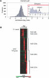
Figure 3. Defining Puf Target RNAs
(A) Distribution of average Cy5/Cy3 fluorescence ratios from four independent microarray hybridizations analyzing Puf3p targets. The arrow depicts enrichment of COX17 mRNA, which is known to bind to Puf3p (Olivas and Parker 2000). The red dashed line indicates the threshold applied for defining 220 target RNAs (a magnification is shown of the enriched region). (B) Cluster of RNA targets for Puf proteins. Rows represent genes (unique cDNA elements) and columns represent individual experimental samples. Each Puf protein and an untagged strain (mock control) were assayed in quadruplicate. The color code indicates enrichments (green–red color scale). The number of mRNAs interacting with each Puf protein is indicated in parentheses. mRNAs clustering with the mock controls were removed as false positives (see Materials and Methods).
Figure 4
Figure 4. Classification of mRNAs Interacting with Puf Proteins
(A) Column charts showing compartmentalization of characterized gene products encoded by the Puf targets. The same compartments are shown for the entire genome in the columns designed “All” (YPD, May 2003). The number of genes represented in the charts is indicated on the top of columns. An asterisk indicates classes with p values of less than 0.001. (B) Fraction of membrane-associated gene products among the Puf targets. We classified the targets by combining both GO and YPD annotations (May 2003). “Plasma membrane” (light blue) is a subpopulation of the total membrane-associated proteins (blue). Soluble cytoplasmic or nuclear proteins were classified as “non-membrane.” “All” refers to the genome-wide compartmentalization of characterized genes, and respective numbers were retrieved from YPD. “Puf2 Top 40” refers to the 40 highest enriched Puf2p targets and equals the total number of Puf1p targets.
Figure 5
Figure 5. Sequence Motifs Interacting with Puf Proteins
(A) Consensus motifs detected within 3′-UTR sequences of Puf3p, Puf4p, and Puf5p target mRNAs. Height of the letters specifies the probability of appearing at the position in the motif. Letters with less than 10% appearance were omitted. Fraction of genes bearing a motif in the 3′-UTR sequence is indicated to the right. Y-helicase proteins are nearly identical in sequence and were excluded from this analysis. (B) Scheme of three-hybrid assay for monitoring RNA–protein interactions in vivo (Bernstein et al. 2002). (C) β-Galactosidase activity for three-hybrid assay. Proteins assayed are indicated on top, RNAs to the left. Abbreviations: pum, pum-HD; cons., consensus motif; UGU/AGA, UGU in consensus sequence mutated to AGA. (D) Activation of HIS3 reporter gene and resistance to 3-aminotriazole (3-AT), a competitive inhibitor of the HIS3 gene product, in a three-hybrid assay (Bernstein et al. 2002).
Figure 6
Figure 6. Localization of Puf Proteins
TAP-tagged Puf proteins were visualized in fixed cells. DNA was costained with 4′,6-diamidino-2-phenylindole dimethylsulfoxide (DAPI).
Figure 7
Figure 7. Gene Expression Profiling of puf3 Mutants
Distribution of average Cy5/Cy3 fluorescence ratios from three independent microarray hybridizations comparing mRNA levels of puf3Δ with wild-type cells grown in minimal media with glycerol. The left frequency axis refers to all genes (black line); the axis to the right refers to Puf3p and Puf4p (control) targets, shown as red and blue lines, respectively. Relative expression levels of the 220 Puf3p mRNA targets in puf3Δ cells were selectively increased compared to all other mRNAs analyzed (p < 10−34), whereas Puf4p targets were not (p > 0.05). Thirty-nine genes involved in aerobic respiration (according to GO annotation and SGD), but not bound by Puf3p, were similarly enriched (p < 5 × 10⁻⁵) in the puf3 mutant as random sets of 39 Puf3p targets (p < 10−6). Likewise, 220 randomly selected mRNAs coding for mitochondrial proteins that were not associated with Puf3p in the experiments herein were weakly enriched in the mutant (p < 10−8).
Figure 8
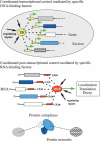
Figure 8. Specific Proteins Bind Functional Groups of Genes for Regulation
At the transcriptional level (top), transcription factors (TFs) regulate initiation of transcription (green arrow) in the nucleus by binding to sequence elements (yellow box) proximal to their target coding regions (boxes). At the post-transcriptional level (middle), RBPs regulate decay, translation, or localization of mRNAs in a coordinated fashion by interaction with sequence/structural elements in the RNA that are often found in 3′-UTR regions (red box). Functional relations at the protein level (bottom) can be reflected at both the transcriptional and post-transcriptional levels: sets of genes that encode functionally related proteins, such as subunits of stochiometric complexes (blue) or components of the same regulatory or metabolic pathway (gray and cross-hatched boxes), may be regulated by common transcription factors and their mRNAs post-transcriptionally coregulated by RBPs (dashed interactions).

References

    1. Albertsen M, Bellahn I, Kramer R, Waffenschmidt S. Localization and function of the yeast multidrug transporter Tpo1p. J Biol Chem. 2003;278:12820–12825. - PubMed
    1. Andersen P, Kedersha N. Stressful initiations. J Cell Sci. 2002;115:3227–3234. - PubMed
    1. Bagnat M, Chang A, Simons K. Plasma membrane proton ATPase Pma1p requires raft association for surface delivery in yeast. Mol Biol Cell. 2001;12:4129–4138. - PMC - PubMed
    1. Bailey TL, Elkan C. Stanford, California; Menlo Park, California: AAAI Press; 1994. Fitting a mixture model by expectation maximization to discover motifs in biopolymers. Proceedings of the Second International Conference on Intelligent Systems for Molecular Biology; August; pp. 28–36. - PubMed
    1. Bernstein DS, Buter N, Stumpf C, Wickens M. Analyzing mRNA–protein complexes using a yeast three-hybrid system. Methods. 2002;26:123–141. - PubMed

Publication types

MeSH terms