. 2001 Aug;13(8):1891-905.
doi: 10.1105/tpc.010102.

G2/M-phase-specific transcription during the plant cell cycle is mediated by c-Myb-like transcription factors

Affiliations

G2/M-phase-specific transcription during the plant cell cycle is mediated by c-Myb-like transcription factors

M Ito et al. Plant Cell. 2001 Aug.

Erratum in

  • Plant Cell 2001 Sep;13(9):2159

Abstract

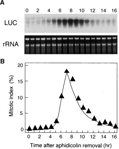
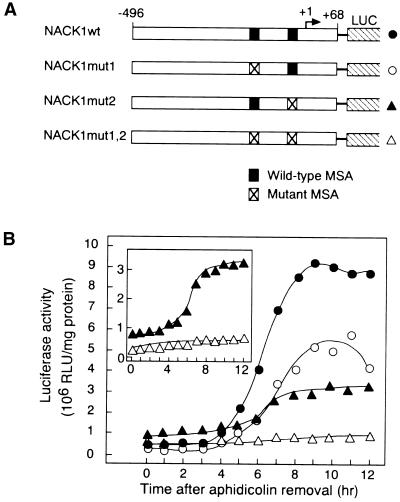
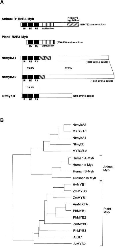
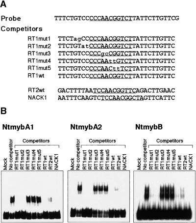
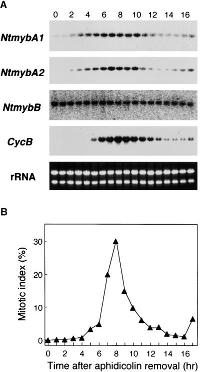
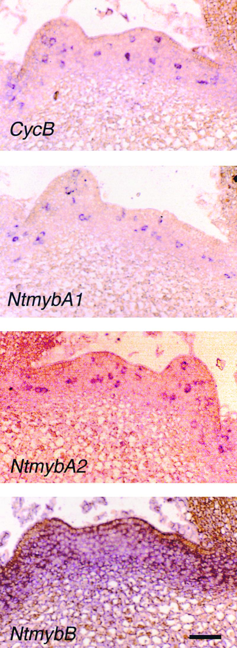
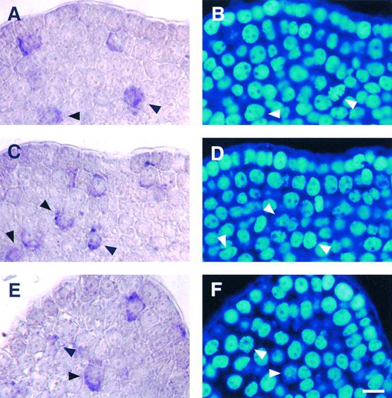
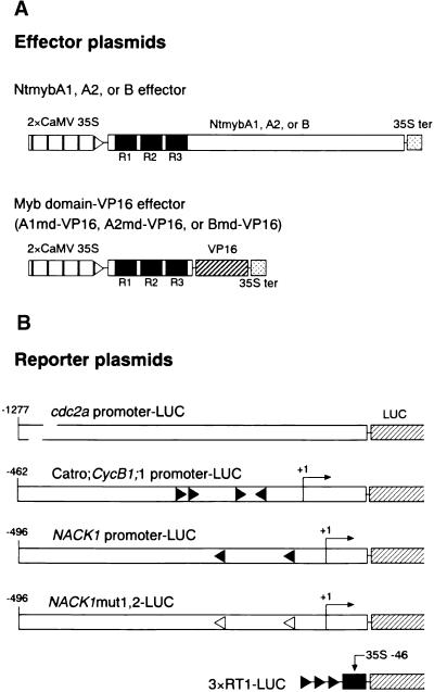
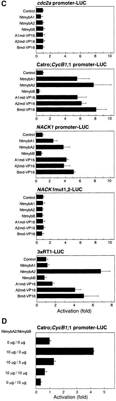
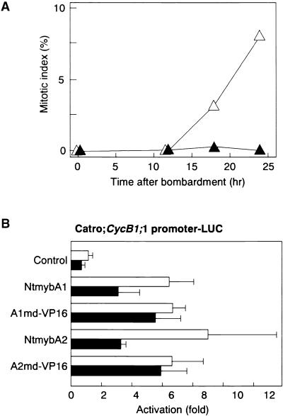
Plant B-type cyclin genes are expressed specifically in late G2- and M-phases during the cell cycle. Their promoters contain a common cis-acting element, called the MSA (M-specific activator) element, that is necessary and sufficient for periodic promoter activation. This motif also is present in the tobacco kinesin-like protein gene NACK1, which is expressed with timing similar to that of B-type cyclin genes. In this study, we show that G2/M-phase-specific activation of the NACK1 promoter also is regulated by the MSA element, suggesting that a defined set of G2/M-phase-specific genes are coregulated by an MSA-mediated mechanism. In a search for MSA binding factors by yeast one-hybrid screening, we identified three different Myb-like proteins that interact specifically with the MSA sequence. Unlike the majority of plant Myb-like proteins, these Myb proteins, NtmybA1, NtmybA2, and NtmybB, have three imperfect repeats in the DNA binding domain, as in animal c-Myb proteins. During the cell cycle, the level of NtmybB mRNA did not change significantly, whereas the levels of NtmybA1 and A2 mRNAs fluctuated and peaked at M-phase, when B-type cyclin genes were maximally induced. In transient expression assays, NtmybA1 and A2 activated the MSA-containing promoters, whereas NtmybB repressed them. Furthermore, expression of NtmybB repressed the transcriptional activation mediated by NtmybA2. Our data show that a group of plant Myb proteins that are structurally similar to animal c-Myb proteins have unexpected roles in G2/M-phase by modulating the expression of B-type cyclin genes and may regulate a suite of coexpressed genes.


Figures

Figure 1.
Figure 1.
G2/M-Phase–Specific Activation of the NACK1 Promoter. (A) G2/M-phase–specific transcription of the NACK1 promoter–LUC transgene in BY2 cells. Tobacco BY2 cells transformed with the NACK1 promoter (−496 to +68)–LUC construct were arrested by aphidicolin treatment. Aphidicolin was removed (0 hr), and cells were harvested at 1-hr intervals. Total RNA was isolated and hybridized with the LUC coding region as a probe. Ethidium bromide staining of the rRNA is shown as well. (B) Change in mitotic index during synchronous cultures. The efficiency of cell synchronization was checked by measuring the mitotic index at 1-hr intervals.
Figure 2.
Figure 2.
G2/M-Phase–Specific Activation of the NACK1 Promoter Is Dependent on the MSA Elements. (A) Scheme of NACK1 promoter–LUC derivatives used for stable transformation of BY2 cells. Numbers indicate nucleotide positions relative to the transcriptional start site (+1). Wild-type and mutant MSA sequences are shown by closed boxes and crossed boxes, respectively. (B) Changes in LUC enzymatic activity during the cell cycle. The transgenic BY2 cells were synchronized by aphidicolin treatment and harvested at 1-hr intervals for LUC assays. Promoter constructs are designated with the symbols shown at right in (A). The inset shows LUC activity of NACK1mut2 and NACK1mut1,2 constructs with an expanded scale. RLU, relative light unit.
Figure 3.
Figure 3.
Structures of NtmybA1, A2, and B Proteins. (A) Scheme showing structures of Myb proteins. Structures of NtmybA1, A2, and B are shown together with functional domains of animal R1R2R3-Myb and typical plant R2R3-Myb proteins. R1, R2, and R3 motifs in DNA binding Myb domains are shown as closed boxes. Repeat sequences found in NtmybA1 are shown as hatched boxes. Amino acid identities within and outside of the Myb domains are shown for NtmybA1, A2, and B. (B) Phylogenetic tree of Myb proteins. The tree was constructed with the UPGMA tree program (Genetyx Software Development Co. Ltd., Tokyo, Japan) based on amino acid sequences of the R2 and R3 motifs in the Myb domains of Ntmyb proteins and other members of the Myb family isolated from plants and animals: c-Myb, A-Myb, and B-Myb from human; Drosophila melanogaster Myb; MYB3R-1, MYB3R-2, AtMYB2, and AtGL1 from Arabidopsis; ZmMYB1, ZmMYB3, and ZmMYBC from maize; PhMYB1, PhMYB2, and PhMYB3 from petunia; AmMIXTA from Antirrhinum majus; and HvMYB1 from barley.
Figure 4.
Figure 4.
Characterization of the DNA Binding Affinity of the Ntmyb Proteins. (A) Nucleotide sequences of oligonucleotides used as probe and competitors in electrophoretic mobility shift assays. Sequences of MSA elements are underlined. (B) Electrophoretic mobility shift assays of Ntmyb proteins. Binding reaction mixtures were incubated with the probe and either mock-translated product (Mock) or in vitro synthesized NtmybA1, A2, and B in the presence or absence of a 40-fold molar excess of unlabeled oligonucleotide competitors.
Figure 5.
Figure 5.
Change in the Levels of Transcripts for NtmybA1, A2, and B during the Cell Cycle. (A) RNA gel blot analysis of NtmybA1, A2, and B. Tobacco BY2 cells were synchronized by aphidicolin treatment. Cells were collected at 1-hr intervals after release from aphidicolin block. Gene-specific probes for NtmybA1, A2, and B were used for hybridization. To detect Nicta;CycB1;3 (CycB) mRNA, full length cDNA was used as a probe. Ethidium bromide staining of the rRNA is shown as well. (B) Change in mitotic index during synchronous cultures. The efficiency of cell synchronization was checked by measuring the mitotic index at 1-hr intervals.
Figure 6.
Figure 6.
Localization of the Transcripts of NtmybA1, A2, and B in Tobacco Shoot Apices. Longitudinal sections of shoot apices were hybridized with digoxigenin-labeled antisense RNA probes of Nicta;CycB1;3 (CycB) and NtmybA1, A2, and B and viewed under a bright field. Hybridization signals are visible as purple color development. Sense RNA probes did not produce any hybridization signals (data not shown). Bar = 50 μm.
Figure 7.
Figure 7.
Periodic Accumulation of NtmybA1 and A2 Transcripts during the Cell Cycle in Tobacco Shoot Apices. Longitudinal sections of shoot apices were hybridized with digoxigenin-labeled antisense RNA probes of Nicta;CycB1;3, NtmybA1, and NtmybA2. They were counterstained with DAPI and viewed with bright field or epifluorescence. Arrowheads indicate mitotic cells with hybridization signals. (A) and (B) Nicta;CycB1;3. (C) and (D) NtmybA1. (E) and (F) NtmybA2. (A), (C), and (E) show transcript localization (bright field). (B), (D), and (F) show DAPI-stained nuclei (epifluorescence). Bar in (F) = 10 μm.
Figure 8.
Figure 8.
NtmybA1 and A2 Function as Activators, and NtmybB Functions as a Repressor. (A) Scheme of effector plasmids used in transient assays. NtmybA1, A2, and B effector plasmids contained the corresponding full-length cDNAs. A1md–VP16, A2md–VP16, and Bmd–VP16 effector plasmids contained fusions between Myb domains from NtmybA1, A2, and B, respectively, and the VP16 activation domain (VP16). All of these constructs were placed between a double CaMV 35S promoter (2×CaMV 35S) and the terminator signal of the CaMV 35S gene (35S ter). R1, R2, and R3 motifs in the Myb domains are shown as black boxes. (B) Scheme of reporter plasmids used in transient assays. Promoters from cdc2a, Catro;CycB1;1, and NACK1 genes were fused to the LUC reporter gene. In NACK1mut1,2–LUC reporter plasmids, both of two MSA elements were mutated. The 3×RT1–LUC reporter contained three tandem copies of the RT1 sequence (a 9-bp MSA motif from the Catro;CycB1;1 promoter) fused to the CaMV 35S basal promoter (positions −46 to +1). Numbers indicate nucleotide positions relative to the transcriptional start site (+1). Arrowheads indicate the positions and orientations of the MSA elements. Black arrowheads, wild-type MSA sequences; white arrowheads, mutant MSA sequences. (C) Promoter activation by NtmybA1 and A2 is dependent on the MSA element. The LUC reporter plasmids cdc2a promoter–LUC, NACK1 promoter–LUC, Catro;CycB1;1 promoter–LUC, NACK1mut1,2–LUC, and 3×RT1–LUC were cotransfected with effector plasmid expressing full- length NtmybA1, A2, and B and Myb domain–VP16 fusions (A1md–VP16, A2md–VP16, and Bmd–VP16) into BY2 protoplasts. As a control, each reporter plasmid was transfected without effectors (Control). (D) NtmybB represses transcriptional activation mediated by NtmybA2. The Catro;CycB1;1 promoter–LUC reporter plasmid was transfected into BY2 protoplasts together with various combinations of effector plasmids expressing NtmybA2 and B. All LUC activities are expressed relative to the reporter construct alone (value set to 1). Error bars indicate ±sd.
Figure 8.
Figure 8.
NtmybA1 and A2 Function as Activators, and NtmybB Functions as a Repressor. (A) Scheme of effector plasmids used in transient assays. NtmybA1, A2, and B effector plasmids contained the corresponding full-length cDNAs. A1md–VP16, A2md–VP16, and Bmd–VP16 effector plasmids contained fusions between Myb domains from NtmybA1, A2, and B, respectively, and the VP16 activation domain (VP16). All of these constructs were placed between a double CaMV 35S promoter (2×CaMV 35S) and the terminator signal of the CaMV 35S gene (35S ter). R1, R2, and R3 motifs in the Myb domains are shown as black boxes. (B) Scheme of reporter plasmids used in transient assays. Promoters from cdc2a, Catro;CycB1;1, and NACK1 genes were fused to the LUC reporter gene. In NACK1mut1,2–LUC reporter plasmids, both of two MSA elements were mutated. The 3×RT1–LUC reporter contained three tandem copies of the RT1 sequence (a 9-bp MSA motif from the Catro;CycB1;1 promoter) fused to the CaMV 35S basal promoter (positions −46 to +1). Numbers indicate nucleotide positions relative to the transcriptional start site (+1). Arrowheads indicate the positions and orientations of the MSA elements. Black arrowheads, wild-type MSA sequences; white arrowheads, mutant MSA sequences. (C) Promoter activation by NtmybA1 and A2 is dependent on the MSA element. The LUC reporter plasmids cdc2a promoter–LUC, NACK1 promoter–LUC, Catro;CycB1;1 promoter–LUC, NACK1mut1,2–LUC, and 3×RT1–LUC were cotransfected with effector plasmid expressing full- length NtmybA1, A2, and B and Myb domain–VP16 fusions (A1md–VP16, A2md–VP16, and Bmd–VP16) into BY2 protoplasts. As a control, each reporter plasmid was transfected without effectors (Control). (D) NtmybB represses transcriptional activation mediated by NtmybA2. The Catro;CycB1;1 promoter–LUC reporter plasmid was transfected into BY2 protoplasts together with various combinations of effector plasmids expressing NtmybA2 and B. All LUC activities are expressed relative to the reporter construct alone (value set to 1). Error bars indicate ±sd.
Figure 9.
Figure 9.
NtmybA1 and A2 Have Promoter Activation Abilities in S-Phase–Arrested Cells. (A) Changes in mitotic index after bombardment. BY2 cells from 8-day-old culture were bombarded with plasmids and cultured in the presence or absence of aphidicolin. Mitotic index was measured at the times indicated after bombardment. Closed triangles, cells cultured with aphidicolin; open triangles, cells cultured without aphidicolin. (B) Particle bombardment assay. As a reporter plasmid, Catro; CycB1;1 promoter–LUC was used. As effector plasmids, constructs expressing full length NtmybA1 and A2 and Myb domain–VP16 fusions (A1md–VP16 and A2md–VP16) were cobombarded. The bombarded cells were cultured in the presence or absence of aphidicolin for 24 hr, and LUC activity was assayed. As a control, the reporter plasmid was transfected without effector plasmids (Control). All LUC activities are expressed relative to the control (value set to 1). Error bars indicate ±sd. Closed bars, cells cultured with aphidicolin; open bars, cells cultured without aphidicolin.

References

    1. Albani, D., Mariconti, L., Ricagno, S., Pitto, L., Moroni, C., Helin, K., and Cella, R. (2000). DcE2F, a functional plant E2F-like transcriptional activator from Daucus carota. J. Biol. Chem. 275, 19258–19267. - PubMed
    1. Althoefer, H., Schkeiffer, A., Wassmann, K., Nordheim, A., and Ammerer, G. (1995). Mcm1 is required to coordinate G2-specific transcription in Saccharomyces cerevisiae. Mol. Cell. Biol. 15, 5917–5928. - PMC - PubMed
    1. Bilang, R., Klöti, A., Schrott, M., and Potrykus, I. (1994). PEG-mediated direct gene transfer and electroporation. In Plant Molecular Biology Manual, S.B. Gelvin and R.A. Schilperoort, eds (Dordrecht, The Netherlands: Kluwer Academic Publishers), pp. A1, 1–16.
    1. Black, A.R., and Azizkhan-Clifford, J. (1999). Regulation of E2F: A family of transcription factors involved in proliferation control. Gene 237, 281–302. - PubMed
    1. Brandeis, M., and Hunt, T. (1996). The proteolysis of mitotic cyclins in mammalian cells persists from the end of mitosis until the onset of S phase. EMBO J. 15, 5280–5289. - PMC - PubMed

Publication types

Substances

LinkOut - more resources