. 2024 Mar 21;390(12):1092-1104.
doi: 10.1056/NEJMoa2307952.

Intrathecal Gene Therapy for Giant Axonal Neuropathy

Collaborators, Affiliations

Intrathecal Gene Therapy for Giant Axonal Neuropathy

Diana X Bharucha-Goebel et al. N Engl J Med. .

Abstract

Background: Giant axonal neuropathy is a rare, autosomal recessive, pediatric, polysymptomatic, neurodegenerative disorder caused by biallelic loss-of-function variants in GAN, the gene encoding gigaxonin.

Methods: We conducted an intrathecal dose-escalation study of scAAV9/JeT-GAN (a self-complementary adeno-associated virus-based gene therapy containing the GAN transgene) in children with giant axonal neuropathy. Safety was the primary end point. The key secondary clinical end point was at least a 95% posterior probability of slowing the rate of change (i.e., slope) in the 32-item Motor Function Measure total percent score at 1 year after treatment, as compared with the pretreatment slope.

Results: One of four intrathecal doses of scAAV9/JeT-GAN was administered to 14 participants - 3.5×10¹³ total vector genomes (vg) (in 2 participants), 1.2×10¹⁴ vg (in 4), 1.8×10¹⁴ vg (in 5), and 3.5×10¹⁴ vg (in 3). During a median observation period of 68.7 months (range, 8.6 to 90.5), of 48 serious adverse events that had occurred, 1 (fever) was possibly related to treatment; 129 of 682 adverse events were possibly related to treatment. The mean pretreatment slope in the total cohort was -7.17 percentage points per year (95% credible interval, -8.36 to -5.97). At 1 year after treatment, posterior mean changes in slope were -0.54 percentage points (95% credible interval, -7.48 to 6.28) with the 3.5×10¹³-vg dose, 3.23 percentage points (95% credible interval, -1.27 to 7.65) with the 1.2×10¹⁴-vg dose, 5.32 percentage points (95% credible interval, 1.07 to 9.57) with the 1.8×10¹⁴-vg dose, and 3.43 percentage points (95% credible interval, -1.89 to 8.82) with the 3.5×10¹⁴-vg dose. The corresponding posterior probabilities for slowing the slope were 44% (95% credible interval, 43 to 44); 92% (95% credible interval, 92 to 93); 99% (95% credible interval, 99 to 99), which was above the efficacy threshold; and 90% (95% credible interval, 89 to 90). Between 6 and 24 months after gene transfer, sensory-nerve action potential amplitudes increased, stopped declining, or became recordable after being absent in 6 participants but remained absent in 8.

Conclusions: Intrathecal gene transfer with scAAV9/JeT-GAN for giant axonal neuropathy was associated with adverse events and resulted in a possible benefit in motor function scores and other measures at some vector doses over a year. Further studies are warranted to determine the safety and efficacy of intrathecal AAV-mediated gene therapy in this disorder. (Funded by the National Institute of Neurological Disorders and Stroke and others; ClinicalTrials.gov number, NCT02362438.).


Figures

Figure 1.
Figure 1.. Electrophysiological Changes after Gene Transfer.
Panel A shows the antidromic median sensory-nerve action potential (SNAP) amplitude from digit II in six participants before and after gene transfer (marked as time 0 on the x axis). Emergence of a previously absent SNAP amplitude response was observed in two participants (at 12 months and 18 months after gene transfer), and an increase in a low but detectable median SNAP amplitude was observed in two participants. Stable ongoing detection of a median SNAP amplitude to at least 6 years after gene transfer was observed in one participant. One participant had a detectable median SNAP amplitude in the natural history study that was subsequently undetectable at baseline and after gene transfer. The remaining eight participants in the study had a persistently absent median SNAP amplitude. The abbreviation vg denotes total vector genomes. Panel B shows the antidromic ulnar SNAP amplitude from digit V in 6 participants. Emergence of a previously absent SNAP amplitude response or an increase in the ulnar SNAP amplitude by 6 to 24 months after gene transfer was observed in five participants. One participant had a persistent and stable ulnar SNAP amplitude to 6 years after gene transfer. The remaining eight participants had a persistently absent ulnar SNAP amplitude. Additional motor responses in the upper and lower extremities and sensory responses in the lower extremity are shown in Figures S8 through S11 in the Supplementary Appendix. A median SNAP amplitude (Panel A) and an ulnar SNAP amplitude (Panel B) of greater than 15 μV were derived at the electromyography laboratory of the National Institutes of Health as the normal value on the basis of data from adults; normal values in children older than 5 years of age have been found to be generally similar to those in adults.
Figure 2 (facing page).
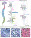
Figure 2 (facing page).. Postmortem Nerve Pathology and Biodistribution 8 Months after Intrathecal Gene Transfer.
Panels A and B show scAAV9/JeT-GAN biodistribution in postmortem nervous-system and peripheral-tissue specimens, respectively, in a participant with giant axonal neuropathy 8 months after receiving the lowest intrathecal dose of scAAV9-JeT-GAN (3.5×10¹³ vg). This participant had undergone spinal fusion surgery and died in the postoperative period after an episode of emesis-induced aspiration followed by anoxemia, which resulted in cardiac arrest and multiorgan failure. Quantitative polymerase-chain-reaction analysis of human codon-optimized gigaxonin (hGANopt) was used to assess vector DNA biodistribution. C denotes cervical, DR dorsal root, DRG dorsal-root ganglia, L lumbar, Mid anatomical middle section along length of given tissue, Prox proximal, SC spinal cord, T thoracic, and VR ventral root. Panels C and D show photomicrographs of DRG samples (1-μm sections stained with toluidine blue; original magnification, 40×) from a participant with giant axonal neuropathy and an age-matched control without the disorder, respectively. Postmortem fixation artifact (evidenced by satellite-cell vacuolation [asterisks]) was present in both samples, but there was no evidence of neuronal loss or inflammation. N denotes neuron (one of several). Panel E shows a photomicrograph of postmortem cervical spinal cord sample (resin-embedded tissue section stained with hematoxylin and eosin; original magnification, 40×) from a study participant with typical giant axonal neuropathy. Numerous giant axons (arrows) were present in the fasciculus gracilis. There was no evidence of inflammatory infiltrates.

References

    1. Opal P GAN-related neurodegeneration. In: Adam MP, ed. GeneReviews. Seattle: University of Washington, 2021. (https://www.ncbi.nlm.nih.gov/books/NBK1136/).
    1. Bomont P, Cavalier L, Blondeau F, et al. The gene encoding gigaxonin, a new member of the cytoskeletal BTB/kelch repeat family, is mutated in giant axonal neuropathy. Nat Genet 2000;26:370–4. - PubMed
    1. Cleveland DW, Yamanaka K, Bomont P. Gigaxonin controls vimentin organization through a tubulin chaperone-independent pathway. Hum Mol Genet 2009;18:1384–94. - PMC - PubMed
    1. Allen E, Ding J, Wang W, et al. Gigaxonin-controlled degradation of MAP1B light chain is critical to neuronal survival. Nature 2005;438:224–8. - PubMed
    1. Bharucha-Goebel DX, Norato G, Saade D, et al. Giant axonal neuropathy: cross-sectional analysis of a large natural history cohort. Brain 2021;144:3239–50. - PMC - PubMed

Publication types

Associated data