. 1997 Feb 10;136(3):545-53.
doi: 10.1083/jcb.136.3.545.

Mdm12p, a component required for mitochondrial inheritance that is conserved between budding and fission yeast

Affiliations

Mdm12p, a component required for mitochondrial inheritance that is conserved between budding and fission yeast

K H Berger et al. J Cell Biol. .

Abstract

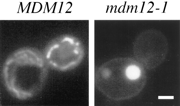
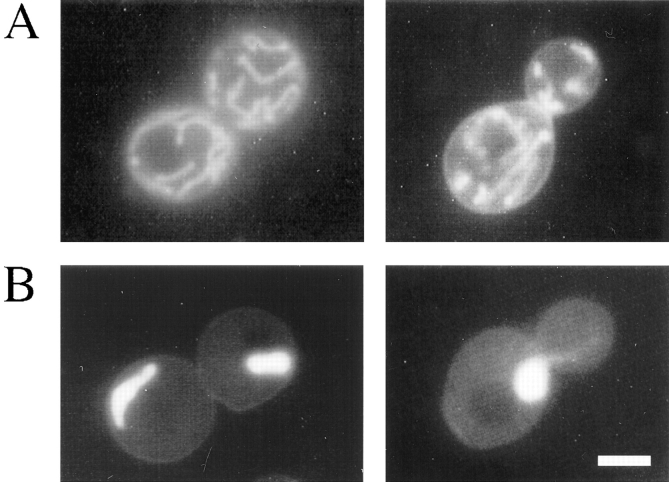
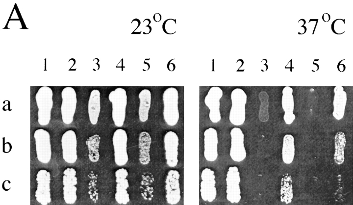
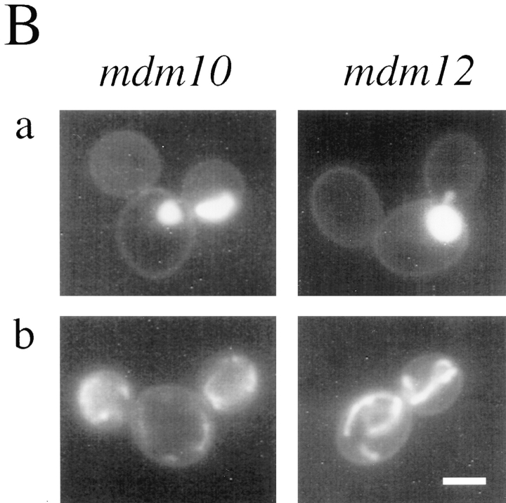
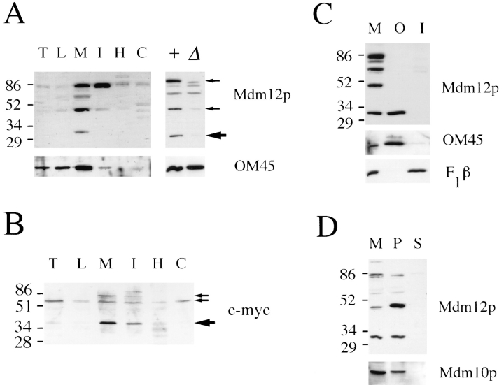
Saccharomyces cerevisiae cells lacking the MDM12 gene product display temperature-sensitive growth and possess abnormally large, round mitochondria that are defective for inheritance by daughter buds. Analysis of the wild-type MDM12 gene revealed its product to be a 31-kD polypeptide that is homologous to a protein of the fission yeast Schizosaccharomyces pombe. When expressed in S. cerevisiae, the S. pombe Mdm12p homolog conferred a dominant-negative phenotype of giant mitochondria and aberrant mitochondrial distribution, suggesting partial functional conservation of Mdm12p activity between budding and fission yeast. The S. cerevisiae Mdm12p was localized by indirect immunofluorescence microscopy and by subcellular fractionation and immunodetection to the mitochondrial outer membrane and displayed biochemical properties of an integral membrane protein. Mdm12p is the third mitochondrial outer membrane protein required for normal mitochondrial morphology and distribution to be identified in S. cerevisiae and the first such mitochondrial component that is conserved between two different species.


Figures

Figure 1
Figure 1
mdm12 mutant cells display giant mitochondria that are defective for distribution to buds. MDM12 (wild type; MYY290) and mdm12-1 (MYY621) cells were grown at 23°C in YPD medium, stained with DASPMI, and viewed by fluorescence microscopy. Representative wild-type (left) and mdm12-1 cells (right) are shown. Bar, 2 μm.
Figure 2
Figure 2
Indirect immunofluorescence confirms the mitochondrial inheritance and morphology defects of the mdm12 mutant. MDM12 (wild type; MYY290) and mdm12-1 (MYY621) cells were grown in YPD medium at 23°C, incubated at 37°C for 2 h, fixed with formaldehyde, and processed for immunofluorescence. Mitochondria were detected with antibodies specific for the mitochondrial outer membrane protein OM14 (B), and mitochondrial and nuclear DNAs were visualized by DAPI staining (C). Brightfield images of the corresponding cells are shown in A. Bar, 2 μm.
Figure 3
Figure 3
Restriction map of MDM12. Represented is a 1,283-bp KpnI–XbaI DNA fragment containing MDM12 complementing activity. The MDM12 open reading frame (solid black arrow) extends from nucleotide 284 to nucleotide 1099. In the mdm12:: URA3 gene replacement, the portion of the MDM12 coding region between the SnaBI and MscI sites, corresponding to most of the MDM12 gene, is replaced with URA3 coding sequences (shaded gray arrow). Restriction sites: H, HindIII; K, KpnI; M, MscI; S, SnaBI; Sp, SphI; X, XbaI.
Figure 4
Figure 4
Alignment of S. cerevisiae Mdm12p with its S. pombe homolog and with a region of Mmm1p. (A) Alignment of S. cerevisiae protein (S.c.) with S. pombe protein (S.p.). (B) Alignment of amino acid residues 123– 178 of Mdm12p with residues 278–333 of Mmm1p. Alignment was performed by the CLUSTALW program (Baylor College of Medicine, Waco, TX). Amino acid identities are indicated by a vertical bar, and similarities are indicated by :. Similarities include R and K, D and E, S and T, N and Q, and A, L, I, M, V, and F. The uncharged region of Mdm12p from amino acids 24 to 40, corresponding to a potential membrane-spanning domain, is underlined.
Figure 5
Figure 5
Expression of the S. pombe Mdm12p homolog in S. cerevisiae produces abnormal mitochondrial morphology. Cells were grown overnight at 23°C in minimal synthetic medium lacking uracil to retain the plasmid and stained with DASPMI. Shown are two different wild-type yeast cells (strain MYY290) harboring either (A) pAC1 (vector) or (B) pAC1 containing mdm12p (plasmid pPM3, encoding the cloned S. pombe Mdm12p homolog). Bar, 2 μm.
Figure 6
Figure 6
SOT1 suppresses growth and mitochondrial morphology defects of mdm10 and mdm12 cells. (A) Dilutions of 10-fold (a), 100-fold (b), and 1,000-fold (c) of strains MYY290 (1), MYY626 (2), MYY503 (3), MYY627 (4), MYY624 (5), and MYY629 (6), were plated onto YPD agar medium and cultured for 80 h at 23°C or 37°C. The relevant genotype of each strain is: (1) MDM10 MDM12 sot1; (2) MDM10 MDM12 SOT1; (3) mdm10 MDM12 sot1; (4) mdm10 MDM12 SOT1; (5) MDM10 mdm12 sot1; (6) MDM10 mdm12 SOT1. (B) mdm10-null (MYY503), mdm12-null (MYY624), mdm10-null SOT1 (MYY627), or mdm12null SOT1 (MYY629) cells were grown overnight in YPD at 23°C and stained with DASPMI. (a) Unsuppressed cells (sot1). (b) Suppressed cells (SOT1). Bar, 2 μm.
Figure 6
Figure 6
SOT1 suppresses growth and mitochondrial morphology defects of mdm10 and mdm12 cells. (A) Dilutions of 10-fold (a), 100-fold (b), and 1,000-fold (c) of strains MYY290 (1), MYY626 (2), MYY503 (3), MYY627 (4), MYY624 (5), and MYY629 (6), were plated onto YPD agar medium and cultured for 80 h at 23°C or 37°C. The relevant genotype of each strain is: (1) MDM10 MDM12 sot1; (2) MDM10 MDM12 SOT1; (3) mdm10 MDM12 sot1; (4) mdm10 MDM12 SOT1; (5) MDM10 mdm12 sot1; (6) MDM10 mdm12 SOT1. (B) mdm10-null (MYY503), mdm12-null (MYY624), mdm10-null SOT1 (MYY627), or mdm12null SOT1 (MYY629) cells were grown overnight in YPD at 23°C and stained with DASPMI. (a) Unsuppressed cells (sot1). (b) Suppressed cells (SOT1). Bar, 2 μm.
Figure 7
Figure 7
Mdm12p is localized to mitochondria by indirect immunofluorescence. Cells were grown in YPD liquid medium at 23°C, fixed with formaldehyde, and processed for indirect immunofluorescence. (A) Wild type (MYY290); (B) mdm12-null SOT1 (MYY629); (C) wild type with SOT1 (MYY626); (D) mdm10-null SOT1 (MYY627). Upper panels show anti-Mdm12p indirect immunofluorescence and lower panels show DAPI staining of nuclear and mitochondrial DNAs. Bar, 2 μm.
Figure 8
Figure 8
Mdm12p is localized to the mitochondrial outer membrane. The mobilities of molecular mass markers, indicated in kD, are shown at the left. (A) Subcellular fractions (left) were isolated from yeast strain MYY626 (wild type with SOT1) grown on semisynthetic lactate medium. Proteins were analyzed by SDSPAGE and Western blotting with antibodies specific for Mdm12p (top) or OM45 (bottom). Large arrow indicates protein species corresponding to the predicted size of Mdm12p, and small arrows indicate two additional species also specific to MDM12. T, total cell homogenate; L, low speed pellet; M, mitochondria; I, intermediate pellet; H, high speed pellet; C, cytosol; +, mitochondrial protein from MDM12 SOT1 cells; Δ, mitochondrial protein from mdm12-null SOT1 cells. (B) Immunoblot analysis of subcellular fractions from wild-type cells (strain MYY290) expressing c-myc–Mdm12p. Fractions are labeled as in A. Large arrow indicates species of predicted size of c-myc–Mdm12p, and small arrows indicate two additional protein species specific to cells harboring c-myc–MDM12. (C) Western blot of wild-type mitochondria (M), purified mitochondrial outer membrane (O), and inner membrane (I) fractions. 20 μg of protein was resolved in each lane. Fractions were analyzed using antibodies to Mdm12p (top), OM45 (middle), and F1β (bottom). (D) Mitochondria (M) were resuspended in 0.1 M Na2CO3. Supernatant (S) and membrane pellet (P) fractions were recovered by centrifugation for 1 h at 100,000 g. Protein samples were analyzed by immunoblotting with antibodies to Mdm12p or Mdm10p.

References

    1. Altschul SF, Gish W, Miller W, Myers EW, Lipman DJ. Basic local alignment search tool. J Mol Biol. 1990;215:403–410. - PubMed
    1. Attardi G, Schatz G. Biogenesis of mitochondria. Annu Rev Cell Biol. 1988;4:289–333. - PubMed
    1. Bairoch A. PROSITE: a dictionary of sites and patterns in proteins. Nucleic Acids Res. 1992;20:2013–2018. - PMC - PubMed
    1. Bereiter-Hahn J. Dimethylaminostyrylmethylpyridiniumiodine (DASPMI) as a fluorescent probe for mitochondria in situ. Biochim Biophys Acta. 1976;423:1–14. - PubMed
    1. Burgess SM, Delannoy M, Jensen RE. MMM1encodes a mitochondrial outer membrane protein essential for establishing and maintaining the structure of yeast mitochondria. J Cell Biol. 1994;126:1375–1391. - PMC - PubMed

Publication types

Associated data