. 1996 Nov 26;93(24):14002-7.
doi: 10.1073/pnas.93.24.14002.

A functional interleukin 12 receptor complex is composed of two beta-type cytokine receptor subunits

Affiliations

A functional interleukin 12 receptor complex is composed of two beta-type cytokine receptor subunits

D H Presky et al. Proc Natl Acad Sci U S A. .

Abstract

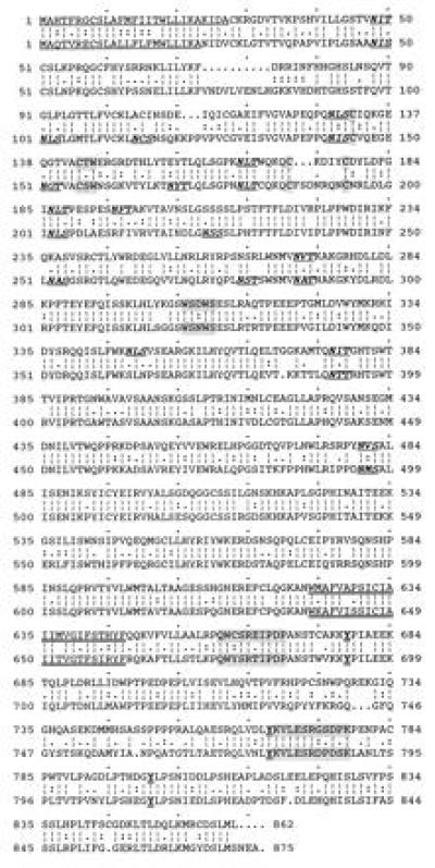
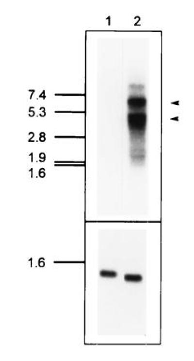
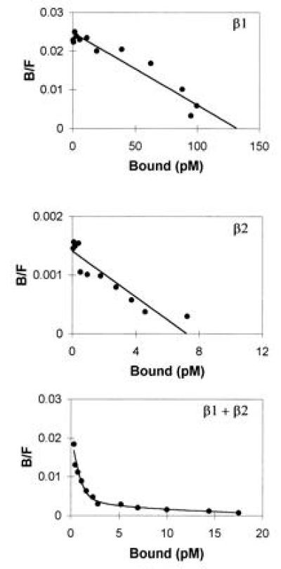
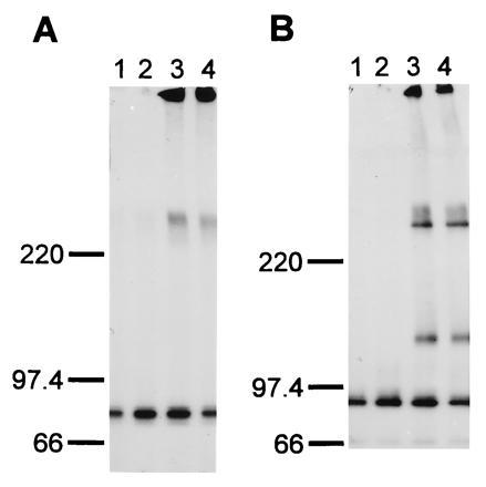
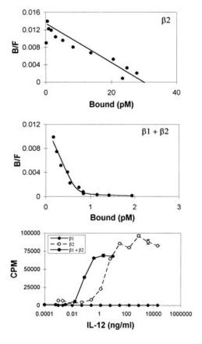
We have identified a cDNA from a human phytohemagglutinin-activated lymphoblast library encoding a protein that binds 125I-labeled human interleukin 12 (125I-huIL-12) with a Kd of about 5 nM when expressed in COS-7 cells. When coexpressed in COS-7 cells with the previously identified IL-12 beta receptor (IL-12R beta) protein, two classes of 125I-huIL-12 binding sites were measured with Kds of about 55 pM and 8 nM, corresponding to the high- and low-affinity binding sites seen on phytohemagglutinin-activated lymphoblasts. This newly identified huIL-12R subunit is a member of the cytokine receptor superfamily, with closest resemblance to the beta-type cytokine receptor gp130 and the receptors for leukemia inhibitory factor and granulocyte colony-stimulating factor. Consequently, we have reclassified the previously identified IL-12R beta subunit as huIL-12R beta 1 and designated the newly identified subunit as huIL-12R beta 2. huIL-12R beta 2 is an 862-amino acid type I transmembrane protein with a 595-amino-acid-long extracellular domain and a cytoplasmic tail of 216 amino acids that contains three tyrosine residues. A cDNA encoding the mouse homolog of the huIL12R beta 2 protein has also been isolated. Human and mouse IL-12R beta 2 proteins show a 68% amino acid sequence identity. When expressed in COS-7 cells, huIL-12R beta 2 exists as a disulfide-linked oligomer with an apparent monomeric molecular weight of 130 kDa. Coexpression of the two identified IL-12R subunits in Ba/F3 cells conferred IL-12 responsiveness, and clones of these cotransfected Ba/F3 cells that grew continuously in the presence of IL-12 were isolated and designated LJM-1 cells. LJM-1 cells exhibited dose-dependent proliferation in response to huIL-12, with an ED50 of about 1 pM huIL-12. Interestingly, Ba/F3 cells transfected with IL-12R beta 2 alone proliferated in response to huIL-12 with an ED50 of about 50 pM, although a role for endogenous mouse IL-12R beta 1 in IL-12 signal transduction in these transfectants cannot be ruled out. These results demonstrate that the functional high-affinity IL-12R is composed of at least two beta-type cytokine receptor subunits, each independently exhibiting a low affinity for IL-12.


Figures

Figure 1
Figure 1
Comparison of aa sequences of human (Upper) and mouse (Lower) IL-12Rβ2 subunits. The gap program (Genetics Computer Group, Madison, WI) was used to construct the alignment. Signal peptides are underlined, potential N-linked glycosylation sites appear in bold italics and underlined, the hallmark sequence/structural motifs characteristic of the cytokine receptor superfamily are shaded, the putative transmembrane regions are doubly underlined, the cytoplasmic box I and II motifs are shaded, and the conserved cytoplasmic tyrosine residues are shown in bold and doubly underlined.
Figure 2
Figure 2
RNA blot analysis of huIL-12Rβ2 expression. (Upper) A blot containing 5 μg of poly(A)+ RNA from uninduced peripheral blood mononuclear cells (lane 1) or PHA-activated (3 days) peripheral blood mononuclear cells (lane 2) was probed with an IL-12Rβ2 cDNA insert. Two major RNAs at 4.5 kb and 6 kb are apparent in lane 2. Exposure time was 16 hr using an intensifying screen at −80°C. (Lower) As a loading control, the blot was rehybridized with a probe for a cell-cycle independent transcript, pHE7 (37). Exposure time was 20 min using an intensifying screen at −80°C.
Figure 3
Figure 3
Coexpression of huIL-12Rβ1 and huIL-12Rβ2 in COS-7 cells yields high affinity binding of 125I-huIL-12. Specific binding of 125I-huIL-12 to COS-7 cells transfected with the indicated expression vectors was determined and analyzed by the method of Scatchard. (Top) huIL-12Rβ1, 6 nM Kd, 123,000 sites per cell; (Middle) huIL-12Rβ2, 5 nM Kd, 6500 sites per cell; (Bottom) both huIL-12Rβ1 and huIL-12Rβ2 expression vectors, 65 pM Kd, 1100 sites per cell and 10 nM Kd, 20,400 sites per cell.
Figure 4
Figure 4
Analysis of huIL-12Rβ2 protein expressed on the surface of transfected COS-7 cells. COS-7 cells were mock-transfected (lane 1) or transfected with a huIL-12Rβ1 expression construct (lane 2), a huIL-12Rβ2-FLAG expression construct (lane 3), or cotransfected with huIL-12Rβ1 and huIL-12Rβ2-FLAG expression constructs (lane 4) as described in Materials and Methods. The cells were metabolically labeled with [35S]cysteine, cell lysates were prepared, and proteins were immunoprecipitated using the M2 anti-FLAG antibody. The precipitated proteins were analyzed on SDS/4-12% polyacrylamide gels under nonreducing (A) and reducing (B; 700 mM 2-mercaptoethanol) conditions. The migration of protein standards electrophoresed in parallel lanes is shown. Exposure time was 6 hr using an intensifying screen at −80°C.
Figure 5
Figure 5
Coexpression of huIL-12Rβ1 and huIL-12Rβ2 in Ba/F3 cells yields high affinity 125I-huIL-12 binding and confers IL-12 responsiveness. Specific binding of 125I-huIL-12 to Ba/F3 cells expressing the indicated proteins was determined and analyzed by the method of Scatchard. (Top) huIL-12Rβ2, 3 nM Kd, 5600 sites per cell; (Middle) huIL-12Rβ1 and huIL-12Rβ2, 60 pM Kd, 120 sites per cell and 17 nM Kd, 500 sites per cell. (Bottom) IL-12-stimulated proliferation of Ba/F3 transfectants expressing huIL-12Rβ1 (♦), huIL-12Rβ2 (○), or both huIL-12Rβ1 and huIL-12Rβ2 (•) was determined as described.

References

    1. Kobayashi M, Fitz L, Ryan M, Hewick R M, Clark S C, Chan S, Loudon R, Sherman F, Perussia B, Trinchieri G. J Exp Med. 1989;170:827–845. - PMC - PubMed
    1. Stern A S, Podlaski F J, Hulmes J D, Pan Y C, Quinn P M, Wolitzky A G, Familletti P C, Stremlo D L, Truitt T, Chizzonite R. Proc Natl Acad Sci USA. 1990;87:6808–6812. - PMC - PubMed
    1. Wolf S F, Temple P A, Kobayashi M, Young D, Dicig M, Lowe L, Dzialo R, Fitz L, Ferenz C, Hewick R M. J Immunol. 1991;146:3074–3081. - PubMed
    1. Gately M K, Desai B B, Wolitzky A G, Quinn P M, Dwyer C M, Podlaski F J, Familletti P C, Sinigaglia F, Chizzonite R, Gubler U. J Immunol. 1991;147:874–882. - PubMed
    1. Chouaib S, Chehimi J, Bani L, Genetet N, Tursz T, Gay F, Trinchieri G, Mami-Chouaib F. Proc Natl Acad Sci USA. 1994;91:12659–12663. - PMC - PubMed

MeSH terms

Associated data