. 1996 Dec 10;93(25):14548-53.
doi: 10.1073/pnas.93.25.14548.

Characterization of serum amyloid A protein mRNA expression and secondary amyloidosis in the domestic duck

Affiliations

Characterization of serum amyloid A protein mRNA expression and secondary amyloidosis in the domestic duck

J T Guo et al. Proc Natl Acad Sci U S A. .

Abstract

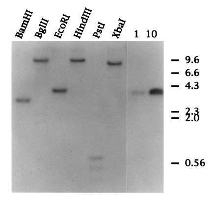
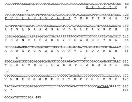
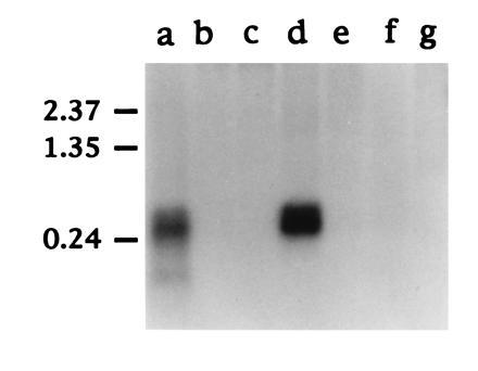
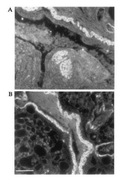
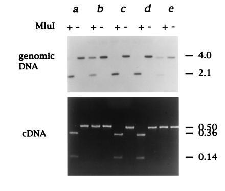
Secondary amyloidosis is a common disease of water fowl and is characterized by the deposition of extracellular fibrils of amyloid A (AA) protein in the liver and certain other organs. Neither the normal role of serum amyloid A (SAA), a major acute phase response protein, nor the causes of secondary amyloidosis are well understood. To investigate a possible genetic contribution to disease susceptibility, we cloned and sequenced SAA cDNA derived from livers of domestic ducks. This revealed that the three C-terminal amino acids of SAA are removed during conversion to insoluble AA fibrils. Analysis of SAA cDNA sequences from several animals identified a distinct genetic dimorphism that may be relevant to susceptibility to secondary amyloid disease. The duck genome contained a single copy of the SAA gene that was expressed in liver and lung tissue of ducklings, even in the absence of induction of acute phase response. Genetic analysis of heterozygotes indicated that only one SAA allele is expressed in livers of adult birds. Immunofluorescence staining of livers from adult ducks displaying early symptoms of amyloidosis revealed what appear to be amyloid deposits within hepatocytes that are expressing unusually high amounts of SAA protein. This observation suggests that intracellular deposition of AA may represent an early event during development of secondary amyloidosis in older birds.


Figures

Figure 2
Figure 2
Southern blot hybridization of duck genomic DNA. Approximately 30 μg of duck genomic DNA was digested overnight with the restriction endonucleases indicated. DNA was resolved on a 1% (wt/vol) agarose gel and transferred to Hybond N membrane (Amersham). The lanes on the right represent 1 and 10 picograms of cloned duck SAA cDNA. The filter was hybridized with a radiolabeled RNA complementary to the 5′ of SAA cDNA (see Materials and Methods).
Figure 1
Figure 1
Nucleotide sequence of duck SAA cDNA aligned with the predicted amino acid sequence. The predicted N-terminal signal peptide and polyadenylylation signal sequence are underlined. The amino acid sequence derived from duck SAA genotype A is shown complete and the six conserved nucleotide changes in type B are shown above.
Figure 3
Figure 3
Schematic of structure and organization of duck SAA gene. Comparison of the duck SAA cDNA and genomic coding sequences. Exons I, II, and III are 146, 140, and 209 nt, respectively; introns are 720 (5′) and 446 (3′) nt, respectively. B, BamHI; E, EcoRI; P, PstI; and M, MluI.
Figure 4
Figure 4
Northern blot hybridization of SAA mRNA. Approximately 1 μg of polyadenylylated RNA from each duck tissue indicated was fractionated on a 1.5% (wt/vol) formaldehyde agarose gel and transferred to nylon membrane (Hybond N; Amersham). The filter was hybridized with a 32P-labeled antisense RNA prepared by in vitro transcription of pGEM.SAA5′ with T7 RNA polymerase (see Materials and Methods). The tissues analyzed were liver (lane a), muscle (lane b), heart (lane c), lung (lane d), intestine (lane e), kidney (lane f), and pancreas (lane g). The position of RNA molecular weight size markers (in kb) are shown on the left.
Figure 5
Figure 5
Expression of AA protein in duck lung tissue. Immunofluorescence staining of ethanol fixed Pekin duck lung tissue with rabbit antisera specific for duck liver AA protein (see Materials and Methods). A and B are two fields on the same section showing strong staining in smooth muscle around the bronchiole. (Bar = ≈200 μm.)
Figure 6
Figure 6
Expression of a single SAA locus in heterozygotes. The SAA genotype was determined by digestion of genomic duck DNA with EcoRI followed by MluI as shown, and SAA DNA detected by Southern hybridization). Type B SAA gene is cleaved by MluI, whereas type A is not (Figs. 1 and 3). SAA gene expression was analyzed by RT-PCR and subsequent MluI restriction endonuclease digestion). Analysis of 5 matched duck liver DNA and RNA samples (a–e) is shown from 15 adult ducks examined in total. Ducks a, c, and d are homozygous for the B allele. Ducks b and e are AB heterozygotes (Upper) but only express the A allele (Lower). The approximate sizes of DNA fragments are shown on the right in kb.
Figure 7
Figure 7
Amyloid deposits in isolated hepatocytes in duck livers at early stage of amyloid disease. (A and B) Photos of the same field on a section cut from ethanol-fixed liver from a duck that exhibited early symptoms of amyloidosis. (A) Rabbit anti-AA fluorescence staining pattern (fluorescein filter). (B) Autofluorescence signal alone (rhodamine filter). (Bar = ≈150 μm.) (C) Three photos of the same specimen obtained using a confocal microscope in which specific AA antibody staining (green) appears colocalized with autofluorescing amyloid deposits (red). (Bar = ≈50 μm.)

References

    1. Rigdon R H. Am J Pathol. 1961;39:369–378. - PMC - PubMed
    1. Cowan D F. Pathol Vet. 1968;5:51–58. - PubMed
    1. Cowan D F. Pathol Vet. 1968;5:59–66. - PubMed
    1. Dougherty E, Rickard C G, Scott M L. Avian Dis. 1963;7:217.
    1. Gorevic P D, Greenwald M, Frangione B, Pras M, Franklin E C. J Immunol. 1977;118:1113–1118. - PubMed

Publication types

Associated data