. 1996 Nov 26;93(24):13931-6.
doi: 10.1073/pnas.93.24.13931.

Promiscuous translocations into immunoglobulin heavy chain switch regions in multiple myeloma

Affiliations

Promiscuous translocations into immunoglobulin heavy chain switch regions in multiple myeloma

P L Bergsagel et al. Proc Natl Acad Sci U S A. .

Abstract

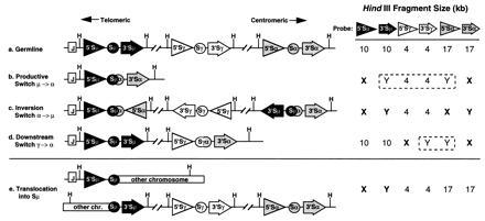
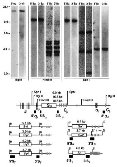
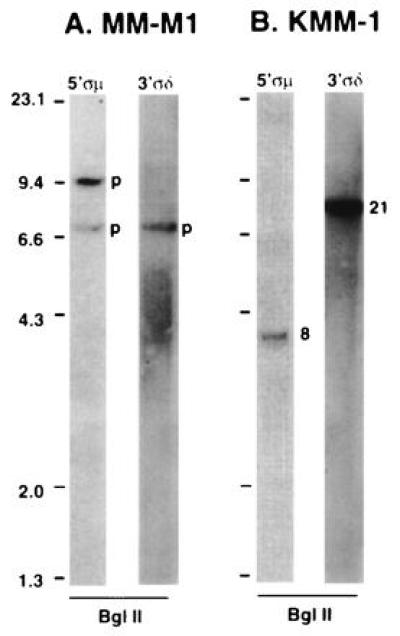
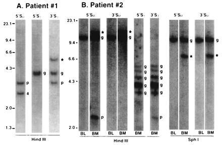
In multiple myeloma, karyotopic 14q32 translocations have been identified at a variable frequency (10-60% in different studies). In the majority of cases, the partner chromosome has not been identified (14q+), and in the remaining cases, a diverse array of chromosomal partners has been implicated, with 11q13 being the most common. We developed a comprehensive Southern blot assay to identify and distinguish different kinds of immunoglobulin heavy chain (IgH) switch recombination events. Illegitimate switch recombination fragments (defined as containing sequences from only one switch region) are potential markers of translocation events into IgH switch regions and were identified in 15 of 21 myeloma cell lines, including seven of eight karyotyped lines that have no detectable 14q32 translocation. From all nine lines or tumor samples analyzed further, cloned illegitimate switch recombination fragments were confirmed to be IgH switch translocation breakpoints. In three of these cases, the translocation breakpoint was shown to be present in the primary tumor. These translocation breakpoints involve six chromosomal loci: 4p16.3 (two lines and the one tumor); 6; 8q24.13; 11q13.3 (in three lines); 16q23.1; and 21q22.1. We suggest that translocations into the IgH locus (i) are frequent (karyotypic 14q32 translocations and/or illegitimate switch recombination fragments are present in primary tumor samples and in 19 of 21 lines that we have analyzed); (ii) occur mainly in switch regions; and (iii) involve a diverse but nonrandom array (i.e., frequently 11q13 or 4p16) of chromosomal partners. This appears to be the most frequent genetic abnormality in multiple myeloma.


Figures

Figure 1
Figure 1
Identification of IgH switch recombination events. A schematic map showing the organization of three IgH switch region loci is shown. The μ (solid), γ (open), and α (shaded) switch region loci are illustrated, with critical elements depicted as follows: circle, switch region; triangle, 5′ switch probe; arrow, 3′ switch probe; and vertical line (H), HindIII restriction enzyme sites. Structural configurations in the germ line and after various recombination events are indicated. The relevant sizes of typical germ-line HindIII restriction fragments detected by each switch probe are shown. The fragments enclosed within dashed lines are deleted during cis recombination but retained during trans recombination. After recombination, germ-line fragments are replaced by rearranged fragments (X and Y) that have different patterns of hybridization with the 5′ and 3′ switch probes, depending on the kind of recombination event. The direction of the triangle and arrow indicates the orientation of transcription, which is unchanged except in the case of inversion.
Figure 2
Figure 2
(Upper) Southern blot analysis of germ-line IgH switch regions. Placental genomic DNA was digested with the indicated restriction enzyme, subjected to electrophoresis on a 0.7% agarose gel, blotted, probed sequentially with various switch probes, and scanned on a PhosphorImager (Molecular Dynamics). The probe is shown above each lane, and lambda HindIII markers are indicated at the left. (Lower) The positions of switch regions (open rectangles), sigma sequences (boxes with horizontal lines), joining region and constant region exons (shaded boxes and lines), 5′ and 3′ switch probes (filled boxes), and restriction sites (S, SphI; B, BglII; H, HindIII) are indicated. The asterisk indicates a SphI site that is present in the 3′ Sα probe and the alpha-2 locus, but not the alpha-1 locus. With the exception of one fragment detected uniquely by the 3′ Sα probe (see text), all 5 pairs of probes cohybridize to the same respective fragment(s).
Figure 3
Figure 3
Southern blot analysis of switch regions in two MM cell lines. Genomic DNA from two cell lines was digested with the indicated restriction enzyme, and Southern blots were analyzed sequentially with various switch probes as described in Fig. 2. The probe is indicated above each lane. (A) MM-M1 cell line. (B) KMM-1 cell line. Illegitimate switch recombination fragments are indicated by 8 and 21 (corresponding to the chromosome involved in the recombination). Productive recombination fragments are indicated by a p.
Figure 4
Figure 4
Southern blot analysis of switch regions in two patients with MM. Genomic DNA from two tumor samples was digested with the indicated restriction enzyme, and Southern blots were analyzed sequentially with various switch probes as described in Fig. 2. The probe is indicated above each lane. (A) Patient 1. (B) Patient 2. For patient 2, DNA was prepared from bone marrow (BM) or blood (BL). Illegitimate switch recombination fragments are indicated by 4 (corresponding to the chromosome involved in the recombination), and ∗ if it has not been cloned. Productive recombination fragments are indicated by a p, and germ-line fragments by a g.

References

    1. Malpas J S, Bergsagel D E, Kyle R. Multiple Myeloma: Biology and Management. Oxford: Oxford Univ. Press; 1995.
    1. Kyle R A. Mayo Clin Proc. 1993;68:26–36. - PubMed
    1. Lai J L, Zandecki M, Mary J Y, Bernardi F, Izydorczyk V, Flactif M, Morel P, Jouet J P, Bauters F, Facon T. Blood. 1995;85:2490–2497. - PubMed
    1. Sawyer J R, Waldron J A, Jagannath S, Barlogie B. Cancer Genet Cytogenet. 1995;82:41–49. - PubMed
    1. Taniwaki M, Nishida K, Takashima T, Nakagawa H, Fujii H, Tamaki T, Shimazaki C, Horiike S, Misawa S, Abe T, Kashima K. Blood. 1994;84:2283–2290. - PubMed