

Associations with photoreceptor thickness measures in the UK Biobank
- PMID: 31857628
- PMCID: PMC6923366
- DOI: 10.1038/s41598-019-55484-1
Associations with photoreceptor thickness measures in the UK Biobank
Abstract
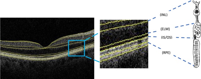
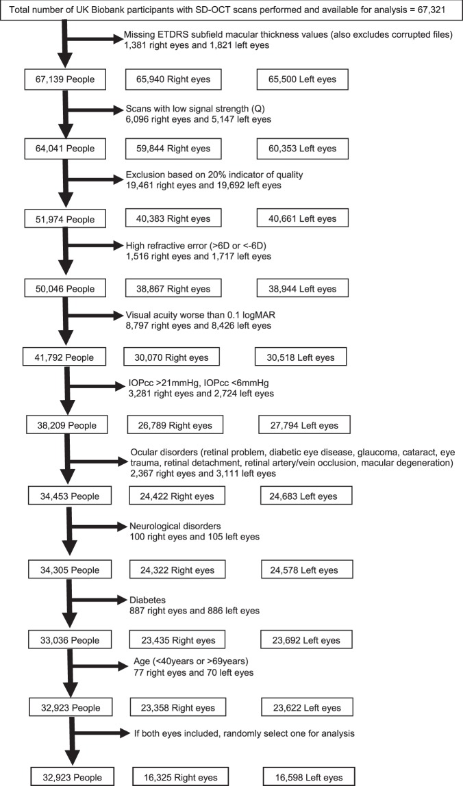
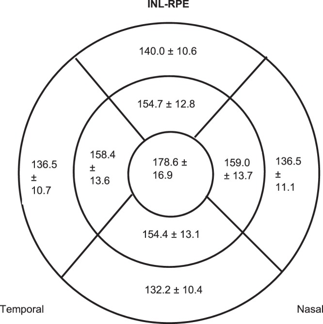
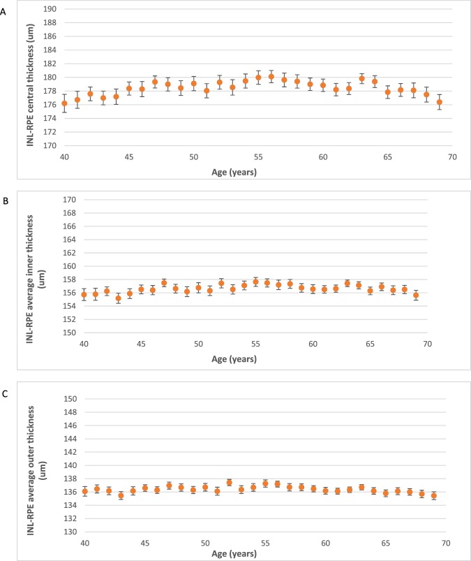
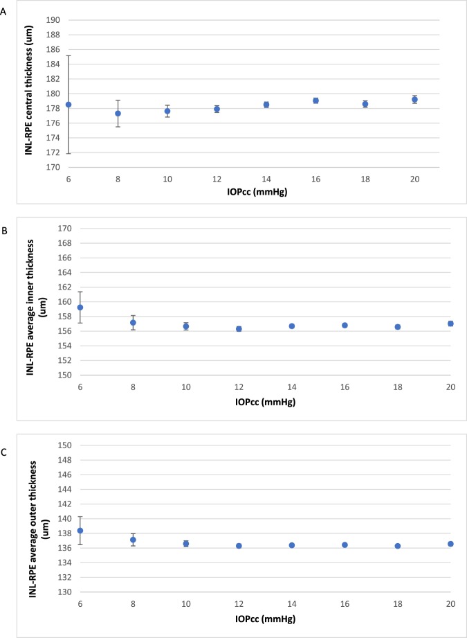
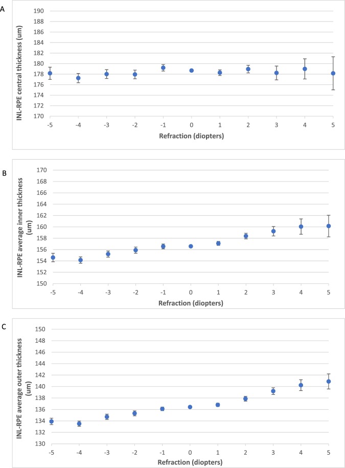
Spectral-domain OCT (SD-OCT) provides high resolution images enabling identification of individual retinal layers. We included 32,923 participants aged 40-69 years old from UK Biobank. Questionnaires, physical examination, and eye examination including SD-OCT imaging were performed. SD OCT measured photoreceptor layer thickness includes photoreceptor layer thickness: inner nuclear layer-retinal pigment epithelium (INL-RPE) and the specific sublayers of the photoreceptor: inner nuclear layer-external limiting membrane (INL-ELM); external limiting membrane-inner segment outer segment (ELM-ISOS); and inner segment outer segment-retinal pigment epithelium (ISOS-RPE). In multivariate regression models, the total average INL-RPE was observed to be thinner in older aged, females, Black ethnicity, smokers, participants with higher systolic blood pressure, more negative refractive error, lower IOPcc and lower corneal hysteresis. The overall INL-ELM, ELM-ISOS and ISOS-RPE thickness was significantly associated with sex and race. Total average of INL-ELM thickness was additionally associated with age and refractive error, while ELM-ISOS was additionally associated with age, smoking status, SBP and refractive error; and ISOS-RPE was additionally associated with smoking status, IOPcc and corneal hysteresis. Hence, we found novel associations of ethnicity, smoking, systolic blood pressure, refraction, IOPcc and corneal hysteresis with photoreceptor thickness.
Conflict of interest statement
S.Y.L.C. and T.A. report no conflict of interest. K.B. reports speaker fees/travel grants/research grants from Novartis, Bayer, Heidelberg, Topcon, Alimera. Q.Y. reports employment by Topcon Medical Systems, Inc. outside the submitted work. P.A.K. reports personal fees from Allergan, personal fees from Topcon, personal fees from Heidelberg Engineering, personal fees from Haag-Streit, personal fees from Novartis, personal fees from Bayer, personal fees from Optos, personal fees from DeepMind, grants from National Institute for Health Research (NIHR), outside the submitted work. C.R. reports employment by Topcon Medical Systems Inc., outside submitted work. P.J.F. reports personal fees from Allergan, Carl Zeiss, Google/DeepMind and Santen, a grant from Alcon, outside the submitted work; P.J.P. reports grants from Topcon Inc, outside the submitted work.
Figures
References
Publication types
MeSH terms
Grants and funding
LinkOut - more resources
Full Text Sources
