Comparative Study
. 1998 Jul 27;142(2):499-509.
doi: 10.1083/jcb.142.2.499.

Bassoon, a novel zinc-finger CAG/glutamine-repeat protein selectively localized at the active zone of presynaptic nerve terminals

Affiliations
Comparative Study

Bassoon, a novel zinc-finger CAG/glutamine-repeat protein selectively localized at the active zone of presynaptic nerve terminals

S tom Dieck et al. J Cell Biol. .

Abstract

The molecular architecture of the cytomatrix of presynaptic nerve terminals is poorly understood. Here we show that Bassoon, a novel protein of >400,000 Mr, is a new component of the presynaptic cytoskeleton. The murine bassoon gene maps to chromosome 9F. A comparison with the corresponding rat cDNA identified 10 exons within its protein-coding region. The Bassoon protein is predicted to contain two double-zinc fingers, several coiled-coil domains, and a stretch of polyglutamines (24 and 11 residues in rat and mouse, respectively). In some human proteins, e.g., Huntingtin, abnormal amplification of such poly-glutamine regions causes late-onset neurodegeneration. Bassoon is highly enriched in synaptic protein preparations. In cultured hippocampal neurons, Bassoon colocalizes with the synaptic vesicle protein synaptophysin and Piccolo, a presynaptic cytomatrix component. At the ultrastructural level, Bassoon is detected in axon terminals of hippocampal neurons where it is highly concentrated in the vicinity of the active zone. Immunogold labeling of synaptosomes revealed that Bassoon is associated with material interspersed between clear synaptic vesicles, and biochemical studies suggest a tight association with cytoskeletal structures. These data indicate that Bassoon is a strong candidate to be involved in cytomatrix organization at the site of neurotransmitter release.


Figures
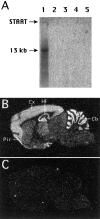
Figure 1
Figure 1
Bassoon transcripts in rat brain. (A) Northern analysis. A Nylon filter containing 20 μg total RNA from brain (lane 1), liver (lane 2), heart (lane 3), and skeletal muscle (lane 4) of 30-d-old rats and from C6 glioma cells (lane 5) was hybridized with 32P-labeled sap7f cDNA insert. (B and C) In situ hybridization. Sagittal sections of rat brain were hybridized with a 40-mer 35S-labeled oligonucleotide probe in the absence (B) or presence (C) of 100-fold excess of unlabeled oligonucleotide. Cb, cerebellum; Cx, cerebral cortex; HF, hippocampal formation; Pir, piriform cortex.
Figure 2
Figure 2
Structure of Bassoon cDNA and gene. (A) Physical map of the rat Bassoon cDNA. Protein-coding region is boxed. Predicted coiled-coil domains are indicated in gray. The extension of analyzed cDNA clones is indicated. Recombined or intron-containing regions are represented by a broken line. (B) Exon–intron organization of the murine bassoon gene. Protein coding region is indicated by filled, 3′-untranslated region by open boxes. Note, intron positions with respect to the open reading frame are indicated in Fig. 3 A. Zn, double zinc-finger motifs; P, heptad repeats, potential phosphorylation sites for proline- directed kinases; poly Q, poly-glutamine stretch.
Figure 3
Figure 3
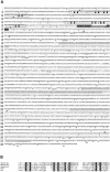
(A) Amino acid sequence of Bassoon. The sequence was deduced from the nucleotide sequence analysis of rat cDNA (upper sequence) and mouse genomic clones (lower sequence). For mouse Bassoon, only amino acid residues that differ from rat Bassoon are printed; dashes indicate deletions. Triangles mark intron positions in the murine bassoon gene, the numbers indicate the position within the reading frame. Zinc- finger motifs are boxed; heptad repeat region is lined in black; coiled-coil domains predicted by the program Macstripe (Lupas et al., 1991) are gray, and the polyglutamine stretch is underlined. The nucleotide sequences are available from EMBL/Genbank, accession nos. Y16563 (rat cDNA) and Y17034, Y17035, Y17036, Y17037, and Y17038 (murine gene). (B) Alignment of the two double zinc-finger motifs of rat Bassoon (Zn-1, Zn-2) with the most closely related zinc fingers of rabphilin-3A (Shirataki et al., 1993), the rabphilin-related protein Noc2 (Kotake et al., 1997) and the Rab3a-interacting molecule Rim (Wang et al., 1997).
Figure 4
Figure 4
Bassoon is highly enriched in synaptic junctional protein preparations. Synaptic proteins were prepared according to Carlin et al. (1980). Western blots (15 μg protein per lane) of the soluble protein fraction (lane 1), the crude membrane fraction P2 (lane 2), the myelin fraction (lane 3), the light membranes fraction (lane 4), the synaptosomal fraction (lane 5), detergent- extracted synaptosomes (lane 6; i.e., One Triton; Kennedy, 1997) and the twice triton–extracted PSD fraction (lanes 7 and 7′; Two Triton; Kennedy, 1997) were probed with mab7f using a chemiluminescent detection system (lane 7′ represents a short exposure of lane 7). Sizes of marker proteins are indicated in kD. Two major protein bands of 420 and 350 kD and several putative proteolytic cleavage products are enriched in the PSD fraction. For comparison, Western blots were reprobed with antibodies against Piccolo, SAP102, synapsin I, and synaptophysin (syph). Note that a major degradation product of synapsin is also detected by the synapsin antiserum (Sikorski et al., 1991).
Figure 5
Figure 5
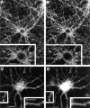
Synaptic localization of Bassoon, synaptophysin, and Piccolo immunoreactivities in mature cultures of hippocampal neurons (21 d in vitro). Double images of hippocampal neurons fluorescently labeled with polyclonal rabbit (A) or mouse (C) antibodies against Bassoon, a monoclonal antibody against synaptophysin (B), or a polyclonal anti-Piccolo antiserum from rabbit (D). Secondary goat anti–rabbit IgG and anti–mouse antibodies were coupled to fluorescein (A), Cy3 (B and C), and Cy2 (D). Insets are close-ups of areas shown in the larger panels. Bassoon co-localizes with the presynaptic proteins synaptophysin and Piccolo (see arrows in C and D). Bars, 10 μm.
Figure 6
Figure 6
Comparison of the distribution of Bassoon and synapsin I immunoreactivities at synapses of the hippocampal CA3 region. Confocal images of the distribution of Bassoon (A) and synapsin I (B). At intermediate magnification the two fluorescent labels appear largely codistributed (C). High magnification images suggest a more restricted localization of Bassoon as compared with synapsin in examples of individual synaptic structures (D–K), including shaft and spine synapses (D–I) as well as mossy fiber terminals (mft, J and K). Dendrites (d) are unstained. Bassoon and synapsin immunoreactivities were detected with Cy2-coupled goat anti–mouse and Cy3-coupled goat anti–rabbit antibodies, respectively. Bars: (A–C) 20 μm; (D–K) 2 μm.
Figure 7
Figure 7
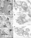
Ultrastructural localization of Bassoon in presynaptic terminals. Electron micrographs of ultrathin sections of rat brain (A–C) and agarose-embedded synaptosomal preparations (D–F) are shown. Analyses were performed with mab7f (A–C) and rabbit polyclonal antiserum against Bassoon (D–E). (A) Excitatory mossy fiber bouton in the stratum lucidum of the hippocampal CA3 region. The presynaptic terminal is densely filled with synaptic vesicles. The peroxidase reaction product is highly concentrated at regions opposite to PSDs (arrowheads). Spiny dendritic processes of pyramidal cells are unstained. (B) Immunopositive axon expansions or terminals contacting a dendritic shaft (d) and spine (s) in the stratum lucidum of CA3. (C) Bassoon immunoreactivity in shaft synapse of the molecular layer of CA3 with apparently separate active zones. Immunoreactivity appears most intense at sites facing PSDs. Bars: (A) 0.5 μm; (B) 0.2 μm; (C) 0.4 μm. (D–F) Ultrastructural localization of Bassoon in nerve terminals by immunogold electron microscopy. Rabbit antibodies were visualized by anti-rabbit IgG coupled with 5 nm gold. Gold particles are highly enriched in presynaptic elements. Bassoon appears not uniformly distributed between all synaptic vesicles (D). Gold labeling of amorphous material between vesicles is preserved, even if the plasma membrane around the presynaptic element is missing (E). Upon incubation without first antibody only very few gold particles are found scattered throughout the preparation (F). PSDs are indicated by arrowheads. Bars (D–F), 0.2 μm.

References

    1. Amaral, D.G., and M.P. Witter. 1994. Hippocampal Formation. In The Rat Nervous System. G. Paxinos, editor. Academic Press Limited, London, United Kingdom. 443–494.
    1. Budnik V, Koh YH, Guan B, Hartmann B, Hough C, Woods D, Gorczyca M. Regulation of synapse structure and function by the Drosophila tumor suppressor gene dlg. Neuron. 1996;17:627–640. - PMC - PubMed
    1. Burns ME, Augustine GJ. Synaptic structure and function: dynamic organization yields architectural precision. Cell. 1995;83:187–194. - PubMed
    1. Carlin RK, Grab DJ, Cohen RS, Siekevitz P. Isolation and characterization of postsynaptic densities from various brain regions: enrichment of different types of postsynaptic densities. J Cell Biol. 1980;86:831–843. - PMC - PubMed
    1. Cases-Langhoff C, Voss B, Garner AM, Appeltauer U, Takei K, Kindler S, Veh RW, De Camilli P, Gundelfinger ED, Garner CC. Piccolo, a novel 420 kDa protein associated with the presynaptic cytomatrix. Eur J Cell Biol. 1996;69:214–223. - PubMed

Publication types

MeSH terms